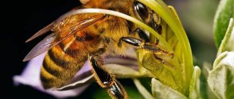
A honey bee is like a biological machine: every part of the insect's body is designed and tuned by nature itself to perform specific tasks. The bee's mouthparts are no exception.
In the lower front part of the Hymenoptera’s head there is a whole enterprise, where there is a real “machine” for grinding, a suction pipe, a device for licking, as well as a small “chemical laboratory” where the nectar undergoes preliminary enzymatic treatment. Let's take a closer look at this wonderful structure.
Bee mouthparts
This is a licking-gnawing apparatus, which means that her mouth simultaneously chews solid food (using the upper jaws for this) and also sucks in liquid food (the lower jaw fuses with the lower lip, together they form a proboscis that sucks in liquid).
The bee's oral apparatus is located below and contains the upper and lower lip, and five soft muscles that contract and thereby expand the enlarged oral cavity.
Covered by the upper lip, the soft protrusion is located below, and muscles also approach it. Under the mouth opening there is a pre-oral cavity. An important part of the bee’s oral apparatus is also the mandibles (upper jaws) - dense chitinous organs with which the bee gnaws solid food.
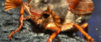
Sometimes it happens that when working with bees, one of them lands on the beekeeper's hand. It does not sting, but a slight pain is felt. This happens because it pulls the hair growing from the skin with its upper (front) jaws, also called mandibles.
The principle of action of RA in bees
The bee's mouthparts are located at the bottom of the head. The insect uses the apparatus in different situations: a complex design is needed for obtaining and processing food, carrying out construction work with wax and wood, cleaning the hive, and assessing the taste of nectar and other substances.
Mechanical restoration
The maxilla RA, also called the mandible, is a paired organ that is used as a cutting and defensive tool.
With the help of mandibles, insects chew pieces of wax, crush wood (during the formation of the entrance), capture and remove garbage and foreign insects from the hive.
Exhaustion
The shape of the mandibles varies among individuals of the bee family.
In the worker bee they are most strongly and well developed. Their chewing edges are rounded; on the upper side there is an expansion and a spoon-shaped depression. Small comb-like ridges are also located here. With the help of the sucking part of the structure, bees manage liquid food: they get nectar from flowers, and also eat honey from honeycombs.
Licking
Using a flat appendage located at the end of the proboscis, the bee can lick nutritional substances from a flat surface. This design feature helps the insect collect food not only by sucking - if the depth is insufficient, a licking device is used.
Taste rating
With the help of taste buds located on the oral appendages, the bee evaluates the chemical composition and taste of the substance.
Chemical treatment
With their upper jaws, worker bees perform a variety of actions: they chew the anthers on the stamens of flowers when collecting pollen, bite off pieces of beebread in the cells, knead wax during the construction of honeycombs, collect propolis, remove the caps from cells with sealed honey, etc.
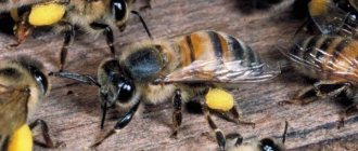
With the help of special enzymes that are secreted by glands internal secretion, the bee pre-processes the nectar. The extracted flower juice is chewed and split, changing the original composition. The prepared substance is already received in the hive by receiving insects, sucking the prey from the forager’s mouth.
Mechanism of operation
The mandibles and upper lip are used to capture prey and roll up pieces of wax or other materials when building a nest. The tongue serves to penetrate the nectaries of flowers. The remaining valves of the maxillae and lower lip are adjacent to the glossae and form a series of channels through which saliva flows down and food flows up. There are disagreements among researchers regarding the details of the mechanism that ensures the movement of liquids.
The drone will also independently gnaw the lid of the cell from which it hatches, because it also has (albeit a very small) maxillary tooth.
An insect has to perform very different actions. Gnaw a tree, expanding the entrance, chew wax, preparing a pulp during construction, and also suck up nectar, reaching for it.
The head has a dense covering, but additionally, chitin penetrates it inside, forming a special structure - the tentorium.
Therefore, the parts of the mouth have reliable support, and the head itself is very strong. Bees use it to compact pollen into cells.
General characteristics of a bee
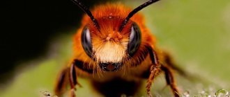
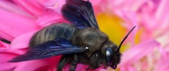
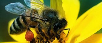
In total, there are about 21 thousand species and 520 genera of bees. They are found on 6 continents. Nature created them so that they are able to feed on nectar and pollen, making them the main pollinators of flower plants. Bees draw in nectar, which is a source of energy, with the help of a long proboscis, and find it and pollen thanks to their antennae. This is their main sense of smell. All individuals of the bee family have one pair of antennae. They consist of segments, the number of which depends on the sex of the individual.
Bees necessarily have two pairs of wings, one pair of which (front) is larger than the second (back). Everyone knows what color a bee is: black with yellow transverse stripes, but the body length varies, depending on the species, from 2.1–39 mm. The smallest size are dwarf bees, the largest are representatives of the species Megachile pluto, living in Indonesia. The bee development cycle has the following stages: egg, larva, pupa, and adult. The egg is laid in a cell.
The type of newborn individual (queen, drone, worker) depends on which part of the queen cell the egg is placed in. The egg laid by the queen is located vertically in the cell. When fertilization occurs, the embryo begins to develop, the position of the egg in the cell will change (it will gradually tilt to the side). After 68–76 hours, required for the development of all organs of the embryo, the egg will lie horizontally at the bottom of the cell.
At this point, the bees place food next to the egg, which will contribute to the destruction of the egg shell and the emergence of a tiny larval worm from it. The bees constantly add food, so the larva is surrounded by food, which is constantly mixed due to the movements of the worm. The hatched larva is only 1.6 mm long and transparent. By the end of the first day of its life, it already reaches 2.6 mm and its body becomes less transparent.
Important! After sealing, the larva of the future worker bee loses weight, while the future queen, on the contrary, gains weight. Because of this, nurse bees gnaw off the lid in advance and nibble the queen’s cocoon to make it easier to leave it.
On the third day of life, the larva will fill the entire bottom of the cell and have a matte color. From this moment on, different larvae begin to be fed differently. Thus, the diet of worker bees from the first day of life consists of milk, then it is replaced with honey and beebread. Future queens are fed exclusively with milk. Drones are also fed milk, but it is even more satisfying. Over the course of 5 (queen), 6 (worker) and 7 (drone) days, the larva molts 4 times. At the end of the last day, nurse bees seal the cells with larvae.
The pupation process begins. How many days this will take also depends on the type of larva. Until the pupa has turned into an adult insect, the molting process occurs a couple more times and after the last shedding of the old shell, the young insect gnaws through the lid and crawls out. In total, it takes 20 days to develop for a worker bee, 16 for a queen bee, and 24 for a drone.
Lower jaw (maxilla)
This is a pair and consists of a main segment (cardo), or pendant, a stem (stipes) and an external saber-shaped blade, or galea. The pendant is movably connected via a condyle to a depression at the edge of the head capsule.
Three pectoral muscles are attached to the stem (with the contraction of which the proboscis moves forward into the working position during the absorption of food) and the internal chewing blade (which serves to regulate the functioning of the food and secretory canals).
The stem is attached to the front end of the pendant. At the external end of the stem there is a rudimentary maxillary palp.
The initial (primary) form in insects is the gnawing type of mouthparts, and all other types, or modifications, originated from it. The bee's mouthparts are of the gnawing-sucking-licking type. The oral apparatus includes the upper and lower lips, paired upper and lower jaws.
The anterior parts closely overlap one another, forming the anterior and lateral sides of the proboscis canal.
During feeding of young larvae of worker bees or the queen, the chewing blades are retracted, thereby opening the preoral cavity and allowing the secretion of the pharyngeal glands to escape.
In the extended position, the galea of the right and left mandibles are folded together, connecting along the rib.
Due to the movement of the chewing blades, a straight channel is formed while sucking nectar with the proboscis.
Mouthparts of a honey bee. The proboscis of bees.
Mouthparts of a honey bee. The proboscis of bees.
The initial (primary) form in insects is the gnawing type of mouthparts (orthopteroid). All other types originated from it - licking, piercing, gnawing, sucking, etc. The mouthparts of bees belong to the licking-gnawing type. The bee chews with its upper jaws, and the lower jaws, together with the lower lip, have formed an elongated proboscis, with which the bee sucks (sucks in) liquid food.
The bee's oral apparatus was studied in detail by Snodgrass (1942). The description and terminology of these organs are given according to his work.
Bee pharynx and esophagus : A - longitudinal section of the head; B - subpharyngeal plate; B - cross section of the beginning of the pharynx (above) and the middle of the pharynx (below) -, - cibarium; k - clypeus; vg - upper lip; ef - epipharynx; p - mouth; l — blade of the hypopharyngeal plate; I am the tongue; c - sa-livarium; in - excretory opening of the lower labial gland; PM - preoral sac; hfp - hypopharyngeal plate; sk - salivary canal; g - pharynx; tm - tentorial occipital bridge; p - esophagus; m - brain; hk - sensitive cells; o - opening of the excretory canal of the pharyngeal gland; mv - rectifier muscle; inter - constrictor muscle; mr - dilator muscles; gs - hypopharyngeal suspensorium; c - suspensor protrusion; mg - muscles of the pharynx; gf - hypopharynx.
In the lower anterior part of the head there is the upper lip (Fig. A, bd), or labrum, a flat, highly sclerotized plate hanging freely under the clypeus. The inner wall, common to the clypeus and upper lip (epipharyngeal surface), is flexible and elastic; it forms the anterior wall of the expanded cavity - the cibarium (c). This cavity is located in the bee immediately behind the mouth opening (p).
Five short muscles are stretched between the clypeus and the anterior wall of the cibarium, the contraction of which expands the cavity of the cibarium (dilator muscles - mr).
Between the lower lip and the mouth opening there is a soft protrusion - epi fa rinke (ef), which is partially covered by the upper lip. Muscles from the clypeus approach the epipharynx; they play a role in the process of swallowing food.
Hypopharynx. Below the oral opening is the preoral (preoral) cavity. A convex tongue-like appendage hangs into it - the hypopharynx (HP). In the anterior part of it there is an oral opening, in the posterior part there is an excretory opening (in) the lower labial gland. The hypopharynx arose from the sternites of the head segments.
The anterior wall of the hypopharynx is formed by the subpharyngeal, or hypopharyngeal, plate (Fig. A, B, gfp). At the anterior end of this plate there is a small flat triangular gipopharyngeal blade (l), which hangs freely into the preoral cavity. On the sides of the subpharyngeal plate there are openings (Fig. B, o) for the excretory canals of the pharyngeal glands. Further back from the plate, a septum extends, separating the anterior part of the pharynx—the cibarium (c) from the pharynx proper (d). At the edges of the subpharyngeal plate there are small thickenings (sclerites), from which two long narrow sclerotized rods extend upward - hypopharyngeal suspensoria (HS). In the place where they approach the upper wall of the pharynx, the suspensories form protrusions (v), to which two pairs of muscles are attached: elongating (mv), or protractors, and retractors (inter), or retractors. When the first muscles contract, the lumen between the cavity of the cibarium and the pharynx opens; when the second muscles contract, the lumen closes.
At the posterior end of the hypopharyngeal plate there are two clusters of sensitive, or sensory, cells (cc).
The process of absorption of liquid food is carried out by expansion and contraction of the pharyngeal pump. This pump consists of two cavities: the anterior one - the cibarium (c) to the expanded part of the pharynx (d). When the cibarium expands under the influence of muscles (mr), fluid is absorbed from the preoral cavity through the open mouth. Then the mouth closes and the numerous muscles (B, mg) located in the wall of the pharynx contract. Under the influence of compression, the liquid moves from the cavity of the cibarium into the wide and then narrow part of the pharynx. With the subsequent expansion of the cibarium, the muscles (B, msg) close the exit to the pharynx and the liquid is again absorbed through the open mouth.
Behind the hypopharyngeal blade there is a pre-oral sac open from the outside (A, pm). In the very posterior part of the hypopharynx there is an elongated cavity of the salivarium (c), at the end of which there is an excretory opening (in) of the lower labial gland. Muscles attached to the walls of the salivarium can open and close the outlet for the secretions of this gland (salivary pump).
The upper jaws (mandibles) are shortened, strong chitinous formations, narrowed in the middle, concave towards the inside (Fig.).
Upper jaws of bees : A - side view B - general view; C - muscles of the upper jaws, D - upper jaws of a worker bee; D - upper jaws of the uterus; B - upper jaws of the drone; HF - upper jaw; vg - upper lip; k - clypeus, c - anterior and posterior articulations of the mandible; t - place of attachment of the mandible; g - groove; b - groove; in - excretory glands; mzh - mandibular gland; kr - root part; rc - incisal edge; p - Mouth; m1 - abductor muscle, m2 - adductor muscle.
Each of the upper jaws (A, hc) is attached to the head capsule at two points far apart from one another. The posterior articulation (A, c) looks like a tubercle, and the anterior one (c) looks like a depression. Between them there is a place of support for the mandible (d). An axis is formed with a point of application at m around which the mandible can move so that its end moves to the side and is brought back.
Not far from the posterior articulation, a muscle (B, m1) is attached to the outer side of the mandible, the contraction of which takes it to the side. The second, stronger adductor muscle (m2) is attached to the inner side of the mandible. Alternate contraction of these muscles leads the mandible to rocking movements.
The touching edges of the mandibles have wide projections and teeth (Fig. D). The inner part of the mandible forms the radical (cr), or molar part; the outer part, equipped with a long and sharp tooth, forms the incisal edge (pk).
Along the inner side of the lower edge of the mandible, from its apex, there is a groove bordered with hairs (B, g). In the middle part of the mandible, this groove turns into a narrow groove (b), which is directed to the excretory opening (c) of the maxillary (mandibular) gland (mg). The secretion of the gland is brought out through this groove and groove.
The bee performs several actions with its mandibles: it tears apart the anthers on flowers when collecting pollen, kneads wax when building combs, chews wood when expanding a very narrow entrance, grabs specks and carries them out of the hive, grabs other bees, enemies and pests. The mandibles tightly enclose and support parts of the proboscis in a folded state and support the unfolded proboscis.
In the queen bee, the mandibles (D) are wider and have a sharp tooth (pk) at the end, with which she chews the cap before leaving the queen cell. The drone's mandibles (E) are underdeveloped and covered with long hairs.
The bee's proboscis is formed by two elongated lower jaws (Fig. B, nch) and an even more elongated lower lip (ng).
Proboscis in bees : A - structure of the lower lip of insects (original type); B - proboscis of a worker bee; nch - lower jaw; ka—cardo; st - stipes; lo - lorum; PM - postmentum; me - mentalum; prm - prementum; pg - paraglossae; u - palps; gl - glosses; I am the tongue; l - spoon; ng - lower lip.
The lower jaw (maxilla) of a bee (pictures above and below) consists of two main segments - cardo (ca) and stipes (st). The bee's cardo is a small thin rod connected through a condyle (A, a) to a depression on the edge of the head capsule. Continuing further from this joint, the cardo forms a process (o), to which a muscle (m4) is attached, starting from the wall of the head capsule. Under the action of the muscle (m4), the cardo, having a fulcrum in the condyle (a), moves the short arm of the lever back and down, as a result of which the long arm straightens forward and upward, pushing the entire lower jaw into the working position.
Stipes (st) is attached to the anterior end of the cardo. It is a long and wide plate. In its original form in insects, stipes bears two maxillary lobes and a palp. The large outer lobe (galea) has a blunt end, the inner one (lacinia) is pointed. The palp (s) usually consists of several segments. The bee also has the same structure of the lower jaw, but individual parts of this organ have changed greatly.
In a bee, a long saber-shaped galea (Fig. below, A, d), pointed at the end, and a rudimentary palp (psh), consisting of two segments, extend from the stipes (st). There are two parts in the galea: a short main one - subgalea (B, sr) and a long one - terminal galea (gk). The subgalea is attached by a wide plane to the stipes, at the end it narrows, bends and passes into the terminal galea. A clearly defined rib (rg) runs along the end part. In the extended position, the galea of the right and left mandibles are folded together, connecting along the rib. The anterior parts of the galea closely overlap one another, forming the anterior and lateral sides of the proboscis canal.
The structure of the lower jaw of a worker bee (according to Snodgrass): A - general view of the lower jaw; B-E - details of the structure of the lower jaw; G - position of the proboscis in a folded (non-working) state; k - clypeus; ka—cardo; cm—stipes; g - galea; l - lacinia; m1-m4 - muscles that straighten the proboscis; rsch - rudimentary palp; ls — lacinial rod; сг - subgalea; rg—galea rib; t - tentorium; gk - terminal galea; I am the tongue; u - palps; p - lever; a — cardo condyle; o - process of cardo; lp - cushion-shaped lobe of lacinia; b — articulation of the lacinial rod with a rod-shaped thickening; y - thickening of the galea wall; m5 - muscle that tightens the lacinial cushion; mb, m7 - muscles that flex the maxillae.
Three large muscles are attached to the stipes (Fig. A). One of them (m1) extends from the clypeus (k) and is attached near the junction of the stipes with the cardo; the other (m2) also originates from the clypeus, but is attached approximately to the middle of the stipes; the third muscle (m3) arises from the tentorium (t) and is attached to the middle of the stipes. When all these muscles contract, the angle formed by the cardo and stipe straightens, as a result of which this part of the proboscis moves forward into the working position.
In addition to the galea, the stipes also has a formation attached to it, which Snodgrass takes to be a homolog of lacinia (l). It consists of the following parts: the lacinia itself (D, l) with hairs at the end, a lever (p), a soft cushion-shaped blade (lacinium cushion - lp) and a rod (ls), going towards the galea in the middle of the cushion. A muscle (E, m5) is attached to the lever, which goes to the posterior end of the stipes; When this muscle contracts, the cushion blade rises and stretches.
The bee straightens its proboscis only when sucking food. The rest of the time the proboscis is folded. The maxillae bend under the influence of two muscles (E, m6, m7), which are attached to a slight thickening of the galea wall (E, y). The end of this thickening is connected to the lacinial rod (D, E, lc). The muscle pulls the thickening back, as a result of which this thickening moves around its articulation (b) with the lacinial rod. In this case, the entire galea bends at the place of its narrowing, turning its end down and back (D).
When the m5 muscle is relaxed (Fig. above, E), the lacinial pillows (Fig. below, A, lp) descend downwards and move far away from the upper lip and hypopharynx. The food channel (PC), formed in the proboscis, turns out to be wide open in its upper part near the mouth. With this position of the oral organs, food cannot be absorbed by the proboscis. But the secretion of the pharyngeal glands, entering through the excretory ducts in the hypopharyngeal plate, turns out to be open. The bee adopts this position of the mouthparts when feeding the queen and drones.
When the M5 muscle contracts (Fig. above, E), the lacinial cushions together with the galea (d) rise upward and press tightly against the epipharynx (Fig. below, B, e) and to each other, closing the food canal. In this position, the food channel of the proboscis turns into a closed tube, and the bee can suck in liquid food through it.
Consequently, lacinia in bees serve to regulate the food and secretory channels. They ensure the creation of a straight channel while the pharyngeal pump is operating when liquid is sucked into the proboscis; if the bee feeds the queen, drone or brood, then the lacinia retracts, opening the pre-oral cavity, allowing the secretion to escape out.
The lower lip (labium) is the most complex. The initial type of its structure in insects is shown in the figure above, A. In the lower lip there is a postmentum (pm) - the main section with which the lip is attached to the head. The postmentum, in turn, is divided into two parts: submentum and mentum.
The terminal section of the prementum (prm) extends from the postmentum, bearing two palps (u) and four lobes: two internal ones - glossae (el) and two external ones - paraglossae (pg).
The bee's lower lip retains all the parts typical of insects, but they have changed greatly.
The lower lip of a bee (Fig. above, B) is attached to the head by means of a submentum, which in a bee is called lorum (lo). Lorum consists of two small thin sticks, located obliquely one in relation to the other and connected at the top. The ends of the lorum are connected to the ends of the cardo(ka) of the lower jaws. Attached to the apex of the lorum is a small triangular segment, the mentum (me). It is followed by a wide and long segment prementum. Long thin palps (u) are attached to the end of the prementum.
Base of the proboscis of a worker bee (according to Snodgrass): A - base of the proboscis with the food canal open (front view); B - base of the proboscis with the food canal closed; B - cross section of the proboscis; cm—stipes; I am the tongue; n — palps of the lower jaw; g - galea; rsch - rudimentary palps; e - epipharynx; lp - lacinial pillow; hpf - hypopharynx; p - mouth; PC - food channel.
The palps consist of four segments. Two small muscles (flexor and extensor) are attached to the palp, starting in the prementum. Next come the elongated outer lobes (paraglossae) and the inner ones (glossae), merged into one organ - the uvula (I).
As already indicated, the lorum tightly covers the mentum on the sides, and its ends are attached to the cardo. Such a close connection of these parts ensures their joint movement: if the cardo moves, then the same movement is transmitted through the lorum and lower lip. When the maxilla extends, under the influence of the same muscles, the lower lip simultaneously moves forward.
The lower lip has muscles that move it independently of the maxillae. From the posterior wall of the head capsule, two long and strong muscles extend to the uvula (Fig. below, D, m1, m2) - labial retractors. They are attached to the anterior part of the prementum and, when contracted, pull back the prementum and the entire lower lip. The presence of the prementum connected to the mandibles causes simultaneous retraction of the maxillae. Consequently, the labial retractors serve to retract the entire proboscis. In addition to the posterior end of the prementum, two more muscles extend from the tentorium. They perform the same role, but independently of the maxillae.
The palp (A, u) is attached to the prementum by means of a small palpiger (pa). The first segment of the palp is greatly elongated, the second is shorter, and the last two are very small (Fig. above, B, y). The lengthening of the entire palp is due to the fact that its function has changed and it has turned from a sensory organ into an integral part of the proboscis - the organ of food intake. The palpi are flat and quite wide; they end at the same level with the galea maxillus. When a bee takes food, the palps form the posterior and partially lateral walls of the large proboscis tube (the anterior and partially lateral walls form the galea of the mandibles, Fig. above, B).
The palp can bend under the influence of a muscle located in the prementum and attached by a long cord to a flexible rod in the lower membranous wall of the palpiger.
Paraglossae (Fig. below, A-B, pg) in the form of short semicircular plates cover the base of the tongue on the right and left sides. But paraglossae can also bend away from the uvula. In the pressed position (A), they ensure the passage of saliva into the uvula channel; when extended (B), they open the direct passage of food through a large tube into the mouth.
The structure of the lower lip of a honey bee (according to Snodgrass): A - base of the proboscis, paraglossae shifted; B - base of the proboscis, paraglossae spread apart; B - base of the proboscis from the back side; G - end of the tongue; D - muscles of the tongue; E - cross section of the proboscis; I am the tongue; kya1, kya2 - uvula channels; pg - paraglossae; u - palpi; pzh - gland ducts; prm - prementum; l - spoon; m1, m2 - muscles that flex the uvula; pa - palpzger; hpf - hypopharyngeal plate; op - main plate; os—axial rod of the tongue; ov - main branch of the rod; gs - flexible palpiger rod; ns - outer wall; fifth - cavity of the tongue; pp—lamina prementum; k - tongue pockets.
The uvula canal (B, kya) is located on the posterior side, and the duct of the lower labial gland (B, pzh) opens behind the hypopharynx (hpf) on the anterior side of the uvula canal. When the paraglossus is in a compressed position, the flowing secretion meets the convex main plate (on) of the uvula on its way and flows around it on the sides, falling into two narrow slits between the uvula and the paraglossae. In this way, the pressed paraglossae ensure the transition of the secretion of the lower labial glands from the front side of the uvula to the back, where the fluid enters its canal.
The bee's tongue (A - B, z) is long, thin, almost round, only slightly flattened in the anteroposterior direction. An axial rod (E, os) runs along the anterior wall of the tongue - a dense elastic cuticular thickening. It forms a small groove (k1) along the back of the tongue, the edges of which are so densely lined with hairs that the groove essentially turns into a small canal. The outer wall of the uvula consists of alternating rings of soft elastic and dense cuticle. The rings of dense cuticle are lined with hairs facing forward towards the end of the tongue.
The ringed structure provides greater mobility and flexibility of the tongue: it can change its length and bend to the sides.
On the back side of the uvula, the cuticle forms another invagination, also in the form of a groove (E, k2). Invagination gives rise to right and left pockets (k), due to which the lumen of the canal can increase. The edges of the groove are lined with dense hairs, so that a second channel of the tongue is formed, larger in diameter than the first. At the end of the tongue, the channel ends, and its walls diverge, forming a triangular spoon (G, l). It is lined along the edge with two belts of strong, straight hairs.
At the base, the tongue is divided into two short branches - right and left. Muscles M1 and M2 are attached to each branch in the middle, originating in the posterior part of the prementum. When only one, for example, the right muscle (D, m1) contracts, it pulls back the right branch, but since this branch is elastic, it only bends and pulls the right side of the rod, causing it to shorten, and this entails a rotation of the entire tongue to the right . The turn to the left is carried out by the same muscle (m2), attached to the left branch of the uvula. The tongue returns to a straight position when both muscles relax due to the elasticity of its walls.
Consequently, the bee's proboscis forms three channels of different diameters. The smallest (capillary) channel is located inside the axial rod of the tongue. A medium-diameter channel is formed by the outer cuticle of the uvula. And finally, the canal of the largest diameter is formed by adding together the galea of the maxilla and palps of the lower lip (Fig. above, B). The secretion of the salivary glands (the salivary canal of the uvula) moves through the first channel to the end of the proboscis. Liquid food passes through the second channel in cases where the bee licks droplets of liquid with a spoon. And finally, the largest diameter of the proboscis channel is used to absorb liquid at close range, when there is a lot of it. In this case, the bee immerses the proboscis into the liquid half its length. The tongue inside the tube moves quickly back and forth during suction, accelerating the flow of liquid.
Saliva passes into the uvula using a salivary pump. This is a narrow cavity located in the posterior side of the hypopharynx a. The secretion from the lower labial gland that gets there is pumped by compression of the walls of the cavity caused by the muscles located here.
Features of the digestive system
The crop is a specific bee organ for carrying and processing nectar into honey. It can stretch and hold up to 90 ml of content, this is with a total bee weight of 110 mg! True, it is usually filled no more than half.
The goiter is located between the mouth and the stomach; it is separated from the latter by a special one-way valve - proventriculus. When squeezed, the stomach is more likely to burst than even a small amount of its contents gets into the goiter.
Another specific feature of the bee’s anatomy is the presence of special rectal glands located at the base of the rectum. The fact is that the bee only eats all winter, without excrement.
Enzymes of the pharyngeal, hypopharyngital gland, developed only in working individuals, invert complex sugars into simple ones. The nectar is gradually processed into honey.
They accumulate in the rectum, and their weight can reach half the total weight of the individual. To protect the contents from unwanted processes, the rectal glands secrete an antiseptic enzyme.
Internal structure
The anatomy of the internal organs of a bee is adapted to their main task - honey production. The internal systems of a bee (sectional side view) are shown in the figure below:
Circulatory system
The bee's circulatory system is not closed, but the blood always circulates in a certain direction. This is achieved thanks to the coordinated work of the heart, aorta, abdominal and dorsal diaphragm. The heart is like a long tube and is located along the back. The chest contains the aorta, which supplies blood to the head. The entire tube is divided into 5 chambers connected through partition valves that allow blood to flow in a certain direction (from the abdomen to the head).
During a minute at rest, the heart pulses 60–70 times. Flight increases the pulsation frequency to 150. The dorsal and abdominal diaphragms are also part of the circulatory system. They control the flow of blood into the body. Blood moves to the limbs, antennae and wings thanks to bubbles located at the base of these parts of the body.
Did you know? When a bee stings, it releases 0.3
–
0.8 mg of poison.
Its quantity will depend on the season and age of the insect. A dose of 0.2 g is lethal to humans. This amount of poison can be obtained from 500 -
1000 sting injections.
Nervous system
It is divided into three sections: central, peripheral and sympathetic. The central one includes the brain and the abdominal chain of nerves, which replaces the spinal cord. The frontal ganglion is the beginning of the sympathetic division, which is responsible for the functioning of the digestive, circulatory and respiratory systems. The brain, as the main hub of the nervous system, contains the bulk of neurons.
The greatest number of them is in the optic lobe and mushroom body. As mentioned earlier, the size of the brain depends on the functions of the bee. In drones it is the largest, but at the same time the worker individual has the most developed sections.
A nerve cell (neuron) is the main structural unit of the nervous system. It has one long process (without branches), transmitting nerve impulses, and a branched process, capable of receiving signals from the unbranched process and other neurons and transmitting it via an electrical signal to its neuron. It turns out that the first process plays the role of an output channel for transmitting information, and the second one plays the role of an input channel.
Respiratory system
The structural features of the insect's respiratory system are such that air enters the body through spiracles, which are holes in the cuticle. Three pairs of spiracles are located on the thoracic region, 6 pairs are on the abdomen. Passing through the spiracles, the air is cleared of dust and collected in bags. This air cannot escape back because the spiracles have valves.
From the bags, air is carried through the trachea through tiny annular branches to all systems of the body. The body leaves the air only through the abdominal spiracles. The large first spiracles are reliably protected by thick hair, but mites can still get into them and cause the development of a disease called acarapidosis.
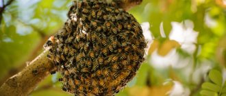
Important! Usually the old queen leaves the family with the swarm, and the young queen remains in the hive.
Reproductive system
There are two types of females in bees: the queen and the worker, but only the first type is capable of producing high-quality offspring. Usually there is only one queen for the entire bee colony. In working individuals, the genitals are highly reproduced. The ovaries and oviducts do not have developed tubes. They begin to develop under certain conditions (the queen died and the worker bees changed their diet).
This will allow the females to produce eggs, but since they do not have a spermatic receptacle, the eggs will be unfertilized and only males can emerge from them. In the uterus, the ovary has approximately 150 tubes, each containing one mature egg. The ovaries and vagina are connected by a paired oviduct.
The spermatic receptacle is connected to the narrow part of the vagina through a canal. The channel acts as a kind of dispenser, which allows several sperm to pass through at the right moment. If fertilization occurs, a worker will emerge from the egg; if not, a drone will emerge.
In section, the reproductive system of the uterus looks like this (1 - ovary, 2 - paired oviduct, 3 - spermatic receptacle, 4 - accessory gland of the spermatic receptacle, 5 - vagina, 6 - reservoir of the large venom gland, 7 - small venom gland, 8 - sting):
Structure and functions of the sting
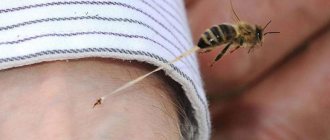
The sting is a means of protection, defense of the bee, and also helps in laying eggs. Only females have it. Looking like a needle, the sting is a modified ovipositor. It is located at the end of the abdomen and covered by its outer segments. Three systems of glands are suitable for it: lubricating, small and large poisonous. The surface of the sting itself is similar to a saw, which allows it to get stuck in the tissues of the enemy.
But, unfortunately, because of this the bee loses it and dies. The longer the sting is in the “victim”, the more poison from the reservoir will enter the body and the more harm it will cause. The poison is a colorless liquid with a special aroma and bitter taste. In air it quickly crystallizes.
Important! The productivity of the uterus is highest in the first year of life and slightly less at the beginning of the second. She is capable of laying about 1500 per season.
–
2000 eggs per day.
The structure of the sting is shown in the figure below, where 1 - sled, 2 - processes of the sled, 3 - oblong plate, 4 - palp, 5 - triangular plate, 6 - stylets, 7 - square plate, M1-M4 - sting muscles, B. Zh. .—large poisonous gland, Yar—reservoir of the poisonous gland, M. Zh.—small poisonous gland:
Bee palps
The palps have lost the function of sensory organs and are part of the proboscis, the organ of food intake. When a bee eats food, the palps form the posterior and partially lateral walls of the large proboscis tube.
Paraglossae cover the base of the tongue on both sides in the form of short semicircular plates that can bend and press the tongue tightly.
In a pressed position, they ensure the passage of salivary gland secretions into the canal of the uvula, and in an extended position, they open the direct passage of food through a large tube into the mouth.
Structure
This type was formed under the influence of the evolutionary relationships of insects with pollinated plants. In the gnawing-licking mouthparts, all the main structures of the original prototype were preserved, but these structures were somewhat modified.
The upper lip and mandibles underwent the least changes. The maxillae and labium became a collection of flattened and elongated elements, and the fused glossae (usually called the uvula) formed into a retractable, groove-like organ.
The bee's tongue consists of alternating rings of soft, elastic and dense cuticle.
Rings of dense cuticle bear hairs facing the end of the tongue. This structure gives the tongue flexibility, strength and elasticity. The rings that form the tongue do not close in one place, due to which a thin capillary channel is formed along the entire length of the tongue.
Structure and functions of the sting
Only females have a sting; drones do not. It developed as a protective adaptation of the family in the process of evolution from the ovipositor and 11–12 abdominal segments.
The sting contains a reservoir of poison - a thick liquid with a sharp-bitter taste, a complex structure, yellowish color and a specific odor. The injection tube consists of two movable slides with inclined serrations.
The notches do not allow the sting to be pulled out from the elastic human skin; when bitten, the entire apparatus breaks off along with the tip of the abdomen. As a result, the bee dies, and the sled tries to penetrate the body, moving alternately, that is, the sting functions on its own, gaining depth and introducing poison.
Therefore, the first aid to a stung person is to carefully remove the part torn from the bee before all the poison is injected.
This is a rather delicate job, since pressing the container will inject the poison under the skin, like from a tube syringe.
So, the structure of the honey bee’s mouthparts differs little from the mouthparts of other insects. The mouthparts of a bee contain all the organs that other insects have; they are partially modified and perform other functions.
Sense organs
It is worth taking a closer look at the sense organs. It is interesting to understand how a bee sees and how its vision works.
Taste
The taste organs include the proboscis. Also, taste buds are located in the pharynx, on the tips of the paws and antennae.
Hearing
Sounds are heard with the whole body and legs. Along the entire outer surface there are special organs that allow you to hear sounds.
Vision
Vision is represented by simple eyes, located at the top of the head in the middle. But to answer the question of how many eyes a bee has, it is necessary to take into account that in addition to simple eyes, bees also have a pair of compound eyes. Each of them perceives its own space. This is due to the fact that they are located on the sides. Then the picture is put together (this is the so-called mosaic vision) and a single picture is formed.
Speaking about what colors they distinguish, it is worth noting that they, first of all, perceive polarized light. This is what allows them to see quite well even with fairly high cloud densities. In all other respects, bees see the same colors as people see.
Smells
Captures odors with its antennae. But it senses surfaces with the help of hairs located on the body. It is these organs that allow you to navigate in the dark. An interesting fact is that drones have 7 times more olfactory receptors, unlike workers.
It is also interesting to know, when examining bees, why they buzz. It is worth noting that these are quite noisy insects with a large body mass (in the world of insects). My wing beats are quite rare, so they set the air in motion, which causes the hum.
The interesting thing is that they can flap their wings without even flying. These are the interesting facts about the structure of this insect that are revealed to the uninitiated.