Life cycle of development of malarial plasmodium in mosquito organs
When sucking the blood of a person with malaria, malarial plasmodia, which are at different stages of development, enter the mosquito’s body, but only gamonts (immature sexual forms) undergo further development. All other plasmodia die. In the stomach of a mosquito, malarial plasmodia travel a complex path.
Rice. 6. The development cycle of malarial plasmodium in the body of a mosquito. Female gamete (17). Formation of the male gamete (18). Fertilization (19). Ookineta (21). Oocyst development (22 and 23). Release of sporozoites from the oocyst (24). Sporozoites in the salivary gland of a malaria mosquito (25).
Maturation of germ cells
In the mosquito's midgut (stomach), gametocytes (immature sexual forms) are converted into gametes (mature sexual forms). Macrogametes or females are formed (mature) from macrogametocytes. Males are formed from microgametocytes. Moreover, from each microgametocyte up to 8 mobile snake-like microgametes are formed. It has been proven that if there are less than 1 - 2 gametocytes in 1 mm3 of the blood of an infected person, mosquito infection does not occur.
Rice. 7. In the stomach of a mosquito, male gamonts “throw out” flagella. The process is called exflagellation.
Fertilization
In 20 minutes. (up to 2 hours) fertilization occurs in the stomach of the mosquito: the microgamete is introduced into the female individual - the macrogamete. When the gametes fuse, they form a zygote. The body of the zygote elongates and it turns into a mobile ookinete. The nuclei of germ cells fuse.
Sporogony
Next, the ookinete penetrates the wall of the mosquito’s stomach, rounds and penetrates its outer wall, becomes covered with a protective membrane, grows and turns into an oocyst. The number of oocysts can be from a few to 500. The entire process from a mosquito bite to the formation of an oocyst lasts about 2 days.
Inside the oocyst, vigorous division of plasmodium nuclei occurs, around which areas of protoplasm condense. The nucleus with a section of protoplasm is called a sporoblast. Inside the sporoblasts, spindle-shaped sporozoites develop, the number of which can reach 10 thousand. The oocyst swells to such a size that the sporozoites float freely in it. The oocysts deposit a pigment, the pattern of which can be used to determine the type of plasmodium.
Rice. 8. The ookinete attaches to the inner wall of the midgut (photo on the left), penetrates it, becomes rounded and penetrates the outer wall, becomes covered with a protective membrane, grows and turns into an oocyst (photo on the right).
Rice. 9. A large number of oocysts on the outer lining of the stomach (a). Opened oocyst and many sporozoites (b). The photo on the right shows oocysts on the outer lining of the stomach.
After the oocyst shell ruptures, the sporozoites enter the body cavity and hemolymph of the mosquito and are spread throughout the body. The largest number of them (hundreds of thousands) accumulates in the salivary glands.
Rice. 10. The photo shows a section of the body of an infected Anopheles mosquito. A huge number of spindle-shaped sporozoites are visible in the hemolymph.
Rice. 11. In the picture on the left there are many sporozoites in the salivary gland of a mosquito. The photo on the right is a view of sporozoites.
After 2 weeks, sporozoites acquire virulence, maintaining infectious properties for up to 2 months. The sporozoites then degenerate.
The timing of sporogony is influenced by the type of mosquito and the ambient temperature.
When mosquitoes are infected with Plasmodium vivax, the insect becomes dangerous after 7 days, Plasmodium falciparum - after 8 - 10 days, Plasmodium malariae - after 30 - 35 days, Plasmodium ovale - after 16 days.
Skin diseases and sexually transmitted diseases and photos
Dermatology (from the Greek derma - skin and logos - science) is the science that studies skin diseases . Dermatology studies the structure and functions of the skin in normal and pathological conditions, the relationship of skin diseases with various pathological conditions of the body, and also clarifies the causes and pathogenesis of various dermatoses, develops issues of diagnosis, therapy, and prevention of skin diseases. Venereology studies the epidemiology, clinical picture, diagnosis, treatment and prevention of sexually transmitted diseases, which include syphilis, gonorrhea, ureaplasmosis, mycoplasmosis, trichomoniasis, chancroid, chlamydia and other human lesions. For this purpose, clinical, histological, microbiological, immunobiological, functional, biochemical, histochemical, experimental and statistical research methods are used. Dermatology is divided into general and private. The subject of general dermatology is general problems (morphology and physiology of normal and diseased skin, general patterns of development of skin diseases, principles of their treatment and prevention, etc.), and private dermatology is concerned with individual dermatoses and syndromes. Dermatology is closely related to the following disciplines: venereology (syphilis, gonorrhea, ureaplasmosis, mycoplasmosis, trichomoniasis, chlamydia), infectious diseases (herpes, AIDS and HIV infection, exanthema and enanthema), pediatrics (rubella, measles, scarlet fever, chickenpox, etc. ), internal diseases (itching, urticaria, etc.), diseases of the endocrine system (myxedema, etc.), surgical diseases (boils, abscesses, etc.), gynecology and obstetrics (dermatoses of pregnant women), neuropathology (leprosy, syringomyelia, etc. .), ophthalmology, psychiatry, etc. Dermatology is of great importance for practical medicine, since skin diseases account for about 10% of all diseases. Skin diseases are in most cases visible to the naked eye. The characteristics of skin diseases, their availability for research in a variety of ways, and the possibility of direct observation of their evolution favor the study of a number of important physiological problems (immunobiological processes, allergies, reactivity, etc.). In its development, dermatology, which became an independent discipline about 100 years ago, has traveled a difficult path. First, naturally, the clinic of skin diseases was studied, then their histomorphology and microbiology; Recently, pathophysiological, pathogenetic and, finally, biological directions have developed, the foundation of which was the achievements of theoretical medicine. The object of study at present is not only a certain dermatosis, but also the background, the “soil” on which it develops, i.e., the condition of a patient suffering from any skin disease, with all its individual characteristics and living conditions. The medical institutions of our country have been significantly replenished with young doctors who need benefits in dermatology and dermatovenereology, which they need in their daily work. Every practicing physician faces the difficult task of understanding numerous skin diseases. Often, even an experienced dermatologist, when examining a patient, cannot quickly make a diagnosis and is forced to turn to reference literature to substantiate his conclusion. It is quite understandable that inexperienced doctors experience much greater difficulties with this. In our opinion, every practicing doctor should always have with him a reference book on dermatology, in which he can find a description of a suspected skin disease, prescribe rational treatment and outline a plan of preventive measures. Considering the wider distribution of certain forms of skin diseases, we considered it appropriate to dwell on these diseases in a little more detail. Particular attention is paid to pyoderma, which is often found among industrial and agricultural workers. This section highlights the classification of pyoderma, describes various clinical forms, their prevention and treatment. In addition to well-known skin diseases, the reference material contains a large number of syndromes, symptom complexes, as well as individual skin diseases , designated by the name of the author. It includes only those syndromes and diseases that have well-defined clinical symptoms and have received general recognition in dermatology. Among infectious diseases, a special place is occupied by skin tuberculosis and leprosy; in this regard, the site outlines the clinical variants of these diseases, their epidemiology and modern methods of therapy. Compared to skin diseases, sexually transmitted diseases (syphilis, gonorrhea, ureaplasmosis, mycoplasmosis, trichomoniasis, chlamydia), which are encountered by doctors of many clinical disciplines, are described in somewhat more detail than skin diseases. To successfully combat these sexually transmitted diseases, doctors are obliged not only to make a correct diagnosis in a timely manner, but also to take an active part in preventive measures. A lot of time has passed since work on the site began. During this time, dermatology received new treatment methods (photochemotherapy, etc.). The number of drugs used in the treatment of skin diseases, various syndromes and symptom complexes has increased. Some drugs have been discontinued. This necessitated a number of corrections and additions. In the proposed material, the sections “Gonorrhea”, “Syphilis”, “Ureaplasmosis”, “Trichomoniasis”, “Mycoplasmosis”, “Chlamydia”, “Herpes”, “AIDS”, “HIV infection”, “Rubella”, “ Measles", "Scarlet fever", "Chickenpox". Skin and venereal diseases are very diverse in their clinical pathology and occur quite often. Therefore, doctors of all clinical specialties often have to deal with patients with skin and venereal diseases. In addition, skin changes often serve only as an external reflection of one or another pathology of internal organs, the central nervous system, or severe systemic diseases. Dermatology and venereology is by no means a “narrow” specialty that only a few “true” dermatologists should master. On the contrary, knowledge of the basics and elements of dermatology and venereology is necessary in the daily work of a doctor of any specialty, including a dentist. The ability to understand these issues should be considered one of the mandatory aspects of professional training, the official duty of a doctor in any clinical specialty. The specifics of teaching dermatovenereology at the Faculty of Dentistry include direct medical guidance, manipulation, prevention and medical examination of patients with diseases of the oral mucosa, which very often are symptoms of many skin diseases and syphilis. Therefore, the main attention should be paid to the study of changes in the oral mucosa during skin and venereal diseases, their clinical varieties, pathogenesis and special treatment. Particular attention should be paid to skin diseases, which often, and sometimes in isolation, appear on the mucous membrane of the mouth and lips. This applies to lichen planus, blistering diseases, exudative erythema multiforme, lupus erythematosus, cheilitis, candidiasis, herpes, etc. A number of diseases in which the mucous membrane of the mouth and lips are affected relatively rarely, for example tuberculosis, leprosy, pustular psoriasis, etc. are offered to future dentists in order to create their understanding of these diseases, without deepening and detailing the symptoms of the disease and treatment. The future dentist must also have an understanding of common skin diseases in order to provide advisory or emergency assistance. These are pyoderma, dermatitis and toxiderma, urticaria, etc. The existing textbooks on dermatovenerology are not sufficiently oriented for teaching skin and venereal diseases at dental faculties and do not fully correspond to the goals and objectives of the training of dentists provided for by the curriculum on skin and venereal diseases. With many skin diseases and syphilis, quite often rashes appear not only on the skin, but simultaneously in the oral cavity, and often skin diseases begin with damage to the oral mucosa, and skin rashes appear much later or the disease manifests itself only as rashes in the mouth or on the red border lips In the latter cases, the main role in diagnosing skin diseases, and sometimes in their treatment and prevention of relapses belongs to dentists. Dermatology as a science was defined in the 18th century. Existed since the 19th century. German, English, French and Russian schools of dermatology at first differed sharply in matters of understanding the essence of skin diseases, but by the beginning of the 20th century. these differences have been smoothed out, and now we can only talk about some features of the direction of scientific research of one or another school, and not about different understandings of the etiology and pathogenesis of diseases. International congresses of dermatologists, dermatological journals, etc. played a huge role in this issue. It should be emphasized that for domestic dermatology, the founders of which were A. G. Polotebnov (1838-1907), A. I. Pospelov (1846-1916), etc., there has always been Characteristic is the attitude towards the patient from the perspective of the whole organism, highlighting the leading role of the nervous system in the pathogenesis of many skin diseases. In the study of the pathology of the oral mucosa, including skin and venereal diseases, the main role belongs to dermatologists, who have described the vast majority of diseases in this area. For example, Setton described deep recurrent aphthae, Turkish dermatologist Behçet - a disease later named after him, Wilson - damage to the oral mucosa with lichen planus, A. I. Pospelov - with psoriasis, Dubrnel - lupus erythematosus on the oral mucosa, Gebra – exudative erythema multiforme, etc. A great contribution was made by Soviet dermatologists, in particular employees of the department of skin and venereal diseases of the Moscow Medical Dental Institute. N. A Semashko. The founder of the dermatostomatological direction was the first head of the department B. M. Pashkov (1899–1973). He belongs to the first description of soft leukoplakia, together with P. D. Sheklakov - benign non-acantholytic pemphigus of the oral mucosa only, the identification of two clinical forms of exfoliative cheilitis - dry and exudative, etc. After B. M. Pashkov, his student A. L became the head of the department Mashkilleyson, who described new forms of precancerous diseases of the lips, meteorological cheilitis, together with T. N. Antonova and E. I. Abramova - vesical-vascular syndrome of the oral mucosa and other diseases. Employees of the department have studied in detail various clinical forms of lichen planus on the oral mucosa and lips, showing the role of gastrointestinal pathology in the pathogenesis of this disease, as well as leukoplakia; various clinical forms of lupus erythematosus on the oral mucosa and red border of the lips are described; classifications of precancerous diseases of the mucous membrane of the mouth and lips, cheilitis have been developed; atopic cheilitis is isolated; the pathogenesis has been studied and treatment for exfoliative cheilitis, Melkersson–Rosenthal syndrome, exudative erythema multiforme has been developed; cystic dermatoses, psoriasis, Behçet's disease, furunculosis, etc. Skin and venereal diseases of adults and children have their own specifics. Due to the significant difference in the reactivity of the body of an adult and a child, in the endocrine status, in the state of the central nervous system, as well as due to the intensive growth of the child’s body and other factors, the same skin diseases often clinically occur with significant differences. In addition, some skin diseases are characteristic mainly or only of adulthood or childhood. For example, occupational skin diseases occur only in adults, epidemic pemphigus, pseudofurunculosis and exfoliative dermatitis - in newborns; Early congenital syphilis manifests itself before the age of 4 years, and late congenital syphilis occurs in the vast majority of cases between 7 and 16–18 years of age. The course of acquired syphilis in children has its own characteristics. These circumstances prompted the creation of separate programs for teaching skin and venereal diseases in the medical and pediatric faculties of medical institutes and textbooks on skin and venereal diseases intended for students of either medical or pediatric faculties. However, a significant number of skin diseases occur in the same way or with slight differences in both adults and children. In addition, practice shows that graduates of the Faculty of Medicine often have to admit children with skin diseases, diagnose and treat them, and graduates of the Faculty of Pediatrics have to do this for adults. Particular attention is paid to protecting the health of mothers and children, steadily improving the quality of medical care, expanding clinical examination, developing new methods and means of prevention, diagnosis and treatment of the most common skin diseases, including dermatoses in children. Skin diseases are an important medical, biological and social problem that requires urgent solutions. It has been established (I.V. Shchutsky) that in recent years the incidence of skin diseases among the child population is 11-12%, and allergic dermatoses are observed in 32% of children under three years of age. 80-95% of children suffering from chronic skin diseases are diagnosed with diseases of the liver, pancreas, and bile ducts, which require special treatment in a hospital setting. Similar results were obtained by N.P. Toropova and co-authors. Skin diseases in children, as a rule, occur against the background of severe pathology of internal organs. Their treatment on an outpatient basis is associated with the release of most mothers from socially useful work in caring for children and with a significant loss of labor resources. The child’s body at different periods of life is distinguished by characteristic anatomical and physiological features, allergic and immunological reactivity. This is due to the influence of hereditary, constitutional, metabolic, neuroendocrine regulatory mechanisms on the formation of the child’s body, including the skin and its functions. The development of skin pathology at different age periods is also significantly influenced by environmental, social and everyday factors, regimen, nutrition, and hygienic care of the child’s skin. Unlike adults, there are some features of the treatment of skin diseases during different periods of childhood. As a result of the constant concern of the Soviet state for the health of mothers and children in our country, in recent years, dermatological offices have been opened at children's clinics, and the network of dermatological departments at children's hospitals and dermatovenerological dispensaries has been expanded. The country's medical institutions have been replenished with young doctors who feel the need for manuals and reference books on pediatric dermatovenerology, which they need in their daily work. The site provides the basic principles of general and local treatment of skin diseases in children. The description of dermatosis ends with recommendations for prevention and clinical monitoring of the child in order to prevent relapses of the disease. At the end of each disease, doses and methods of use of medications most commonly used in pediatric dermatology are given. When performing such labor-intensive and multifaceted work, it was impossible to avoid some shortcomings. In this regard, comments and suggestions from readers will help improve the quality of the material presented in further work on it and will be received with gratitude.
Life cycle of Plasmodium falciparum in humans: exoerythrocytic (preclinical) stage of malaria
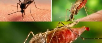
Infection
When an infected female mosquito bites a person, malarial plasmodia at the sporozoite stage enter the human bloodstream with the saliva of the insect. Within 10 to 30 minutes, sporozoites move freely in the blood plasma and then settle in the liver cells. Some of the sporozoites (bradysporozoites) Plasmodium ovale and Plasmodium vivax hibernate, while another part of them, as well as Plasmodium falciparum and Plasmodium malariae (tachysporozoites) begin hepatic schizogony immediately.
Rice. 12. Tissue exoerythrocytic schizogony. 2 - trophozoite, 3 - schizont, 4 - release of merozoites from liver cells into the blood.
Period of tissue schizogony
In liver cells (hepatocytes), the transformation of sporozoites into tissue schizonts occurs, which after 6–15 days divide to form many tissue merozoites. From one sporozoite, from 10 to 50 thousand hepatic merozoites (schizonts) are formed, which enter the blood after 1 - 6 weeks.
When infected liver cells are destroyed, tissue merozoites are released into the blood. This ends the incubation period of malaria and begins the period of erythrocyte schizogony - the period of clinical manifestations.
Hibernation process
Some sporozoites (hypnozoites) of Plasmodium ovale and Plasmodium vivax, once in hepatocytes, turn into inactive forms and hibernate. Parasites can remain in this state for months and years, causing distant relapses.
Rice. 13. Tissue schizont in the liver.
Life cycle of Plasmodium falciparum in humans: erythrocyte (clinical) stage of malaria
After the liver cells rupture, merozoites enter the blood and invade red blood cells. The erythrocyte (clinical) stage of schizogony begins.
Rice. 14. Erythrocyte schizogony. 5 and 6 are ring-shaped trophozoites. 7, 8 and 9 - young, immature and mature schizonts. 10 - erythrocyte merozoites.
Attachment to red blood cells
Attachment of merozoites to the membrane of erythrocytes and invagination into their membranes occurs due to the presence of special receptors on the surface of red blood cells. It is believed that the receptors on the surface of erythrocytes that serve as targets for merozoites are different for different types of Plasmodium.
Rice. 15. Red blood cells infected with Plasmodium vivax (the causative agent of tertian malaria) and Plasmodium ovale (the causative agent of tertian malaria) become enlarged, discolored and deformed, and toxic granularity appears in them. When infected with Plasmodium malariae (the causative agents of four-day malaria) and Plasmodium falciparum (the causative agents of tropical malaria), the shape and size of red blood cells do not change.
Erythrocyte schizogony
Having penetrated red blood cells, schizonts absorb the protein globin (a component of hemoglobin), grow and multiply.
In the erythrocyte, the parasite goes through 4 stages of development:
- Ring (trophozoid) stage.
- Stage of amoeboid schizont.
- Morula (fragmentation) stage. At this stage, the nuclei of schizonts are repeatedly divided (into 6 - 25 parts), and areas of the cytoplasm are separated around them. Erythrocyte merozoites are formed.
- Some parasites go through the stage of gametocyte formation.
A parasite that has penetrated an erythrocyte is called a trophozoid (schizont). It gradually increases in size. A vacuole appears near its nucleus and the trophozoid takes the form of a ring (ring) - a ring-shaped schizont.
As they grow (schizonts feed on hemoglobin), they increase in size and take on the appearance of an amoeba - an amoeba-shaped schizont.
Then, as the schizont grows, it becomes rounded and its nucleus divides many times—the morula stage. Each type of plasmodia has a certain number of nuclei: 12 - 12 in P. vivax, 6 - 12 in P. malariae and P. ovale, 12 - 24 in P. falciparum. This is how erythrocyte merozoites are formed. From the 1st schizont, 8–24 blood merozoites are formed, from which asexual and sexual forms of parasites further develop.
The duration of the erythrocyte schizogony phase is 72 hours in P. malariae, and 48 hours in other Plasmodium species.
During growth, pigment accumulates in the cytoplasm of parasites. Its appearance is associated with the processes of hemoglobin assimilation. Hemoglobin accumulations look like rods or grains. Its color ranges from golden yellow to dark brown.
Rice. 16. During growth, pigment accumulates in the cytoplasm of parasites.
After the destruction of erythrocytes, merozoites enter the blood, some of which re-enter the erythrocytes, others undergo a cycle of gametogony - transformation into immature germ cells, gamonts.
Together with merozoites, heme (the second component of hemoglobin) enters the blood. Heme is a powerful poison and causes acute attacks of malarial fever.
Cycles of erythrocyte schizogony are repeated every 3 days, in other types of malarial plasmodia - every 2 days.
Rice. 17. Destruction of the erythrocyte and release of merozoites into the blood.
Rice. 18. A merozoite of Plasmodium vivax (the causative agent of tertian malaria) that has penetrated into an erythrocyte (thin blood smear).
Rice. 19. Young trophozoids of Plasmodium falciparum (the causative agent of tropical malaria) under a microscope.
Rice. 20. Ring-shaped trophozoites of Plasmodium vivax, the causative agents of tertian malaria (ring stage).
Rice. 21. The photo shows an amoeboid schizont Plasmodium vivax (stage of an amoeboid schizont).
Rice. 22. The photo shows mature Plasmodium vivax schizonts (morula or fragmentation stage).
When red blood cells are destroyed and merozoites are released into the plasma, febrile attacks and anemia develop. When liver cells are destroyed, hepatitis develops.
Tropical diseases.
The site includes basic information about the etiology, pathogenesis, clinical picture, diagnosis, treatment and prevention of protozoal, bacterial, viral and fungal diseases common in hot countries. In addition, data is presented on the hygiene of hot countries and anti-epidemic measures to combat infectious diseases in countries with a tropical climate. Teaching and personal experience in medical institutions in some countries of Africa and Asia were the basis for the creation of this textbook on the main sections of the course of tropical medicine. Tropical diseases are based on sections of the official program for foreign students studying at medical institutes and universities of the CIS countries; the main sections of the course of tropical medicine are considered here. When writing the textbook, the authors did not pretend to be an exhaustive presentation of all nosological forms of diseases found in countries with tropical and subtropical climate, but they meant. The site covers selected issues of tropical medicine, which included protozoal and helminthic infestations, viral infections, and tropical mycoses. The proposed material reflects particularly dangerous infections (smallpox, cholera, plague) and diseases caused by poisonous animals. The greatest share is occupied by clinical issues, diagnosis and treatment of the actual tropical forms of parasitic and vector-borne diseases and mycosis. When creating this section of the site, we used the data accumulated to date in domestic and foreign literature, as well as personal experience in epidemiology, clinical presentation, diagnosis, and treatment of diseases characteristic of subtropical and tropical climate zones. One of the important issues, the presentation of which requires unification, is the nomenclature of chemotherapeutic drugs.
Medical news in the world:
| Is coffee harmful or beneficial? Regular coffee drinkers do not experience an increase in blood pressure after caffeine administration. In irregular coffee drinkers, blood pressure increases after taking both caffeine and coffee or CBC; therefore, it can be assumed that other ingredients in coffee, rather than caffeine, are responsible for this increase in BP. Read more… | Pregnancy – no AIDS! Scientists have concluded that highly active antiretroviral therapy (HART) significantly reduces the risk of transmitting HIV infection to children (transmission of infection to children from mothers who received ART was observed in only 1% of cases), and also helps today to lead a normal, comfortable lifestyle . Read more… | Raw milk can cause serious pregnancy complications A chemical compound found in unpasteurized food was found at unusually high levels in the red blood cells of pregnant women diagnosed with preeclampsia, a serious pregnancy complication that can lead to heart problems. Read more… | Fish oil can be harmful Fish oil supplements that help some heart patients may worsen the condition of others, reports EurekAlert, citing a study from St. Michael and the University of Toronto, Canada, published in yesterday's issue of the Journal of the Canadian Medical Association. Read more… |
| Treatment for impotence has been found!!! Recently, scientists managed to isolate a substance that enhances erection, but acts differently than Viagra (and similar drugs) from spider venom. Read more… | New methods of dental treatment. Toothache let's say: “NO!” ...the discovery of this gene will help recreate damaged tooth enamel, facilitate dental treatment, and even grow new teeth to replace lost ones. Read more… | Curing cancer in 60 minutes is a reality. CyberKnife works with submillimeter resolution, allowing tumors to be clearly localized and eliminated so that the loss of surrounding healthy tissue is minimal - up to 0.1–0.3 mm. Read more… | Karakurt or Black Widow is already in our area. The Black Widow's venom is stronger than that of one of the most dangerous snakes - the rattlesnake (15 times stronger). Read more… |