Parasitic and vector-borne diseases continue to occupy a significant place in human parasitology [1]. Arthropods (phylum Arthropoda
) is one of the most numerous groups of animals, including the class insects (
Insecta
). Contact with certain species of arthropods can cause a number of diseases. Thus, infection with fly larvae leads to the development of myiasis. In Russia, cases of diseases caused by arthropods are quite rare, which makes their diagnosis and timely treatment difficult [2].
Myiasis _
, units
number; Greek, myia
- fly) - diseases of humans and animals caused by the larvae of non-blood-sucking dipterous insects - flies [3].
More often than others, the human body is parasitized by the larvae of synanthropic species of flies of the families Muscidae, Calliphoridae, Sarcophagidae
(order
Diptera
, suborder
Cyclorrhapha
), related in their ecology to humans, and gadflies - grazing species of flies, obligate parasites of warm-blooded animals (families
Gasterophilidae, Oestridae, Hypodermatidae
).
The first descriptions of human myiasis on the territory of our country are given in the works of I.A. Porchinsky (1874, 1875, 1885, 1913), Ya.N. Sokolova (1895), K.G. Samson (1895), H.E. Kusheva (1896), F.O. Yavetsky (1904) [4]. So, I.A. Porchinsky established the role of various types of flies in the occurrence of myiases, studied the specifics of reproduction of a number of species (egg laying, viviparity of larvae), and described the stages of development of larvae. I.A. Porchinsky studied and described in detail the morphology of adults and larvae of flies of the family Sarcophagidae
and primarily
Wohlfartia magnifica
(wohlfart fly), which causes obligate myiases, as well as gadflies. Works by I.A. Porchinsky [5] laid the foundation for a detailed study by entomologists of the development cycle of many species of synanthropic and coprobiont flies.
Geographically, myiases are distributed in tropical climates in the southern United States, Africa, Asia and Australia [3]. In our country, isolated cases of cutaneous myiasis have been described, as they are very rare. Thus, the literature [6] describes a case of migratory subcutaneous myiasis with systemic manifestations in a Russian tourist traveling in Chukotka, which was caused by the larvae of the gadfly Hypoderma
bovis
(cured with ivermectin).
Myiases caused by parasitism of fly larvae are diverse in localization, pathogenesis, and species composition of flies. The generalization and analysis of published materials allowed V.N. Beklemishev to classify myiases and subdivide them into facultative, obligate rapidly developing, obligate slowly developing and myiases caused by botfly larvae. Myiases were subsequently subdivided into random, facultative and obligate [5]. Obligate means that the larvae develop in the body of a human or warm-blooded animal. In case of facultative - on rotting organic remains (meat, vegetables), in the cavities of the ear, nose of a person or animal [2, 7]. Attracted by the smell of rotting tissue, females can lay eggs on open wounds, ulcers, in purulent eyes, ears and become facultative pathogens of ophthalmomyasis, pharyngorinomyasis, otomiasis (auricular), superficial myiasis (cutaneous and subcutaneous). The term “accidental myiasis” is also used, in which the larvae enter the human body accidentally through food. According to localization, random myiases can be intestinal, urinary, or superficial [5]. Genital myiasis has been described in a woman who was unconscious in the forest [8]. In most cases, myiases in humans are caused by parasitizing fly larvae under the skin.
Based on the nature and severity of the course, superficial ( m y iasis
cutis superficialis
) and deep (
m y iasis cutis profunda
) skin myiasis are distinguished.
In superficial cases, the larvae develop in wounds and ulcers; Its causative agents are mainly larvae of flies of the family Calliphoridae
, house flies (
Musca domestica L
), etc. Deep myiases are characterized by a more severe course and are found mainly in tropical countries.
Among them are African (cordylobiasis) and South American (dermatobiasis). They can also be caused by the larvae of flies Wohlfortica magnifica
and a number of others [2].
A parasitic disease in humans is caused by Dermatobia
hominis
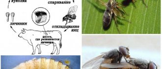
. A parasitic infection caused by large non-blood-sucking arthropod botflies is observed in persons returning from areas of Central and South America. Insect eggs are laid in the skin and then penetrate into the subcutaneous tissue, where the larvae reach maturity. The larva lives in an erythematous papule or nodule, which can be mistaken for an inflamed cyst. The papule has a central pinhole measuring 1-2 mm, which represents the respiratory tube of the larva. Many patients experience discomfort and movement in the skin. The life cycle of such insects is unique in that the female glues her eggs to the abdomen of a mosquito or tick, which involuntarily lays larvae on human skin during a bite or feeding. After maturation, the larva emerges from the body and falls to the ground, and then turns into an adult insect [9].
For the first time, we observed a unique clinical case of epidermal cutaneous myiasis in a 2-year-old girl in Eastern Siberia (Krasnoyarsk Territory). To date, there is no data in the literature describing such dermatoses in the above-mentioned territory. 08/17/16 they brought girl E
. 2 years. According to the mother, rashes on the scalp appeared about 1 week ago; they were accompanied by itching and caused anxiety in the child. The child's mother extracted the larvae from the discharge of the rash and brought it with her on a napkin. During the summer of 2016, the girl and her family repeatedly vacationed in nature in the Ilansky district of the Krasnoyarsk Territory near lakes. All family members are healthy.
From the life history: a child from the fourth pregnancy (one medical abortion, three births), third birth at 32 weeks. The pregnancy proceeded without complications. Premature birth occurred due to rupture of membranes, prolapse of umbilical cord loops, and bacterial vaginitis. The anhydrous period was 9 days. A female fetus with a body weight of 1700 g and a height of 42 cm, a head circumference of 28 cm, a chest circumference of 25 cm, with an Apgar score of 5-7 points was extracted by caesarean section. The child's condition at birth was assessed as severe (weak cry, low muscle tone, weak motor activity, chin tremor), and multiple stigmata of dysembryogenesis were noted. She was transferred to an interdistrict children's hospital with a diagnosis of “prematurity 32 weeks, intrauterine growth retardation of hypotrophic type I degree (15%), Down syndrome.” The condition upon transfer is closer to serious. The subcutaneous fat layer is underdeveloped, stigmata, hypotonia, and hyporeflexia are noted. Ultrasound of the brain revealed immaturity of the brain; X-ray of the chest organs shows the heart and lungs without pathology. Consulted with an ophthalmologist - no pathology was identified. The girl was discharged from the interdistrict hospital under the supervision of a pediatrician.
Past illnesses in the child: in April and May 2015 and in June 2016, acute nasopharyngitis of moderate severity, in December 2015 - acute nasopharyngitis, croup of 0-1 severity; in August 2016, iron deficiency anemia of moderate severity.
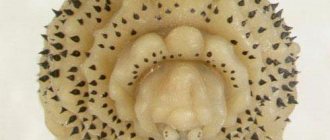
Local status: the pathological process is inflammatory in nature, localized on the skin of the scalp (parietal, occipital, right temporal region); presented by local erythema, nodules with black dots in the center, papulopustules, purulent crusts (Fig. 1). From the holes, larvae are extracted, 3-5 mm in length, ovoid in shape, with a funnel-shaped mouth of a dark color, the body of the larvae is yellowish in color with spines (Fig. 2).
Rice. 1. Child 2 years old. On the skin of the scalp there are areas of erythema, nodules with black dots in the center, pustules, purulent crusts.
Rice. 2. Dermatobiahominis larva, extracted from nodules on the scalp of a 2-year-old child.
A diagnosis was made: myiasis of the scalp, complicated by a secondary infection.
On 08/18/16, in a surgical hospital under short-term general anesthesia (mask anesthesia), surgical sanitation of the lesions was performed, treatment with alcohol-containing antiseptics, and an aseptic bandage was applied. Before sanitation, in order to more easily remove the larvae, an occlusive bandage with baneocin ointment was applied to the scalp for 3 hours. Due to the presence of a local inflammatory reaction, amoxicillin 125 mg 3 times a day was prescribed (the child’s body weight was 10 kg); dressings with potassium permanganate solution, baneocin ointment. In a detailed blood test dated 08/18/16: leukocytes 6.4 109/l, erythrocytes 5.2 1012/l, hemoglobin 91 g/l, granulocytes 44.5%, monocytes 8%, lymphocytes 47.5%, ESR 8 mm/h. The pathological process on the skin completely resolved within 4 days against the background of the above measures. With diagnosed grade I anemia, she was referred for consultation to a local pediatrician.
Thus, clinical and laboratory diagnosis of diseases caused by arthropods is difficult due to the rarity of this pathology. Our experience shows that not only tourists traveling to tropical countries, but also those traveling in the Russian Federation, including Eastern Siberia, should be informed about measures to prevent bites of blood-sucking insects. Staying young children, especially those who are somatically weakened, with congenital malformations and chromosomal abnormalities, near lakes in natural areas can be especially dangerous.
Guzey T.N. — orcid.org/0000−0001−7745−7744
The authors declare no conflict of interest.
How dangerous are gadflies for humans?
Males do not bite - their mouth is adapted only for collecting and consuming flower nectar. Female mammals need the blood of mammals to breed offspring - only fertilized individuals bite. They inject a special substance into the wound that blocks the flow of blood from it, and lays eggs in it. Usually cattle become the incubator for its offspring. Humans are very rare. This can be explained by severe pain during the bite. The female pierces the skin and tries to climb into the wound to lay eggs. This process does not take a second, the bitten person has time to react and drive away the fly biting him. The only exception is the abundance of gadflies, which are difficult to fight off, and an unconscious state (sleep or severe intoxication).
For humans, a gadfly bite is dangerous due to an acute allergic reaction, infection of the wounds and parasitism of larvae in them. The latter develop for several weeks, after which they leave the body through the initial puncture. All this time they give the wearer a lot of unpleasant sensations. Even if the gadfly simply bit, without having time to lay eggs in the wound, the latter is very disturbing.
The resulting itching inevitably leads to scratching. In a hot and humid climate, even the smallest wound is a favorable environment for thousands of microbes. In addition, gadflies are carriers of tick-borne encephalitis, anthrax, polio and a host of infectious diseases.
How to get rid of gadfly larvae?
The consequences of infection with a gadfly larva are treated by an infectious disease specialist. A visual examination alone is not enough to confirm the diagnosis. An enzyme immunoassay blood test is prescribed. If the test is negative, treatment is carried out as for a gadfly bite. If it is positive, specialized specialists are involved, depending on the location of the parasites. Usually this is a gastroenterologist, otolaryngologist, or ophthalmologist. The course of treatment includes the following activities.
- Antibiotic therapy. Drugs are selected individually based on medical history. Along with antibiotics, antiparasitic agents are prescribed.
- Removing larvae. They are neutralized with medications or a scalpel.
With any treatment option, the bite site remains a source of infection throughout its entire period. It must be carefully and regularly treated with an antiseptic with an anti-inflammatory effect. The best option is a solution of Rivanol 0.1%, which combines disinfecting and anti-inflammatory properties.
How to prevent a gadfly bite?
It is impossible to be 100% protected from a gadfly bite in its habitat. The risk of encountering it is highest near farms and grazing livestock. The following recommendations will help to significantly reduce the likelihood of encountering these large biting insects.
- Maximum closed clothing near bodies of water. It is impossible to follow this rule on a river or lake beach in the heat. Horseflies are very active near water. Closed equipment is suitable for mushroom pickers, hunters and lovers of country walks.
- Only shod feet. In summer, on the beach, this is, again, impossible. When fishing or hiking, exposed areas of the legs are not allowed. Socks in the areas between sneakers (sneakers) and trousers will not help - horse flies land and sting everything that moves. They cannot handle only dense, hard materials.
- Avoid walking in tall grass or near grazing livestock. Domestic and wild animals are also attractive to gadflies, just like humans. In the meadow it is better to wear long trousers and rubber boots.
- Swimming areas. Choose only beaches approved by the Ministry of Emergency Situations and Rospotrebnadzor. When the beach season opens, public beaches are checked not only for the presence of parasites in the water.
- Using repellents. Formulations containing at least 50% diethyltoluamide are especially effective against horse flies.
- Rivanol 0.1% to take with you. It does not take up much space, but will allow you to quickly and efficiently treat the wound, preventing itching, swelling and further infection.
- In nature, you can protect yourself by treating the area with a pre-prepared solution of ammonia, lemon juice and a mouthwash with a strong strong odor. All they need to do is spray the picnic area. Don’t be lazy – prepare this solution in advance.
- The aroma of pine needles, wormwood, and tansy repels parasites.
Bodfly bites are very painful, especially if they are not isolated. They cause subsequent inflammatory and infectious diseases, as well as acute allergic reactions. In case of infection with larvae, the consequences are much more serious. Only the bite itself, the pain and itching from it, are immediately noticeable. Allergies usually appear immediately. The consequences of infection may make themselves felt later - the length of the incubation period matters. 2-3 weeks after the bite, it is no longer possible to establish the true cause of the infection, as well as the factor that provoked the exacerbation of a chronic disease. For this reason, we recommend not to neglect preventive measures and first aid for gadfly bites and consult a doctor promptly in case of swelling and obvious intoxication. Keep a bottle of Rivanol 0.1% in your home and car first aid kit, a universal antiseptic with a pronounced anti-inflammatory and antipruritic effect. It will help out in any situation - immediately after a bite, in the treatment of scratching, suppuration and parasite infestation.
Appearance of larvae and what they are
The subcutaneous botfly is a large fly approximately 1.3–1.8 cm long. It has a yellow head with large black eyes, a blue belly, orange legs, and transparent wings. The entire body is covered with hairs, which makes the insect look like a bumblebee. The adult does not feed, using the nutrients accumulated by the larva.
Life cycle
The gadfly is an insect that has a closed chain of transformations. The complete development cycle involves the path from the larva to the adult stage. The insect lives from 3 to 20 days. By the end of life, it loses approximately 1/3 of its own body weight. Under unfavorable climatic conditions, it seems to freeze, living on plants. Any life cycles within the insect's body will be slowed down.
Stages of larval development
The gadfly larva will go through 3 stages of formation inside the human body. At all stages it has a characteristic shape:
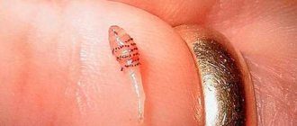
- At stage 1, it looks like a small headless and legless white worm. At the end of the body there is a thickening with 3 black stripes. This stage of formation lasts 7 days, after which it molts and moves on to the next one.
- At stage 2, the larva is large in size and bottle-shaped. After 18 days, the insect molts and moves to the next stage.
- At stage 3, the gadfly will increase in size. After about a month, it will become an adult and will continue to stay in the host’s body for up to 10 weeks, then it will crawl to the surface of the skin and leave the person, falling to the ground.
Each stage is characterized by small black dots and spikes surrounding the thorax.
Important! The larva will feed on the tissues and fluids of the host’s body, dissolving solid components with dermatolytic special enzymes.
What harm can the larvae under the skin cause?
The larva of the gadfly contributes to the development of dermatobiasis, which is an obligate myiasis. This disease is characterized by the formation of nodes under the skin, near the worm that has dug into them, which fester and become inflamed.
The place where the parasite has entered is similar to a mosquito bite. Over time, this area becomes inflamed, irritated and painful. The diameter of the subcutaneous formation reaches 3 cm; its shape is similar to a boil, from which pus is released.
It is worth noting that the larva can invade any part of the human body - the foot, upper limb and even the head. But most often it lives in the legs, armpits and back.
In some cases, the larva settles under the mucous membrane of the eye, resulting in ophthalmomyasis, which often ends in complete loss of vision. This condition is accompanied by the following symptoms:
- redness;
- lacrimation;
- painful sensations.
Also, the larva of the gadfly can live in the nose. In this case, the patient experiences a headache, discomfort in the nose, his sense of smell worsens and the septum is destroyed. At the same time, the mucous membrane swells, and worms can crawl out of the nose on their own.
In addition, the larvae sometimes parasitize the mucous membranes of the lips, mammary glands and penis. If multiple invasions occur, the formations spread over a large area of skin.
After 12 weeks, the larvae in the human body mature, crawl out of it and pupate.
Diagnostics
It is possible to determine that parasites have entered the skin of humans and animals not only by the symptoms and reactions of the infected organism described above. Although an experienced dermatologist or infectious disease specialist will immediately make the correct diagnosis upon examination. If there is any doubt, a special blood test is performed, which shows the amount of antibodies. Are they exceeding the norm? Then it is necessary to begin pest control as quickly as possible.
Allergy to gadfly sting
Another of the most common consequences is an allergy to the bite of a gadfly. Its saliva is one of the strong allergens. An aggressive reaction to it occurs quite often, even in people who do not suffer from other types of allergies. In its absence, swelling, itching and redness disappear after a few days.
The above symptoms are common to all people. When bitten by a gadfly, the allergy gives an additional reaction, manifested in the following.
- Swelling around the bite site.
- Swelling of the eyelids, lips, tongue, larynx, limbs and/or face in general.
- Increased body temperature.
- Hardening of the swollen area.
- Weakness, dizziness, decreased blood pressure.
- Choking, asthmatic attacks, wheezing.
- Nausea, diarrhea, sometimes vomiting.
If any of the above symptoms occur, you should immediately take an antihistamine and consult a doctor. An allergy to a gadfly bite may not appear immediately - it all depends on the dose and nature of the allergen that has entered the blood. It manifests itself especially sharply and quickly in people who already have a history of an allergic reaction to something.
What symptoms does the parasite cause?
The larva can penetrate any part of the human body: arms, legs, chest, head. However, she most often chooses the legs, back and armpits. Sometimes the larvae settle in the eyes and nose.
If gadfly larvae enter a person’s body, then at first no signs can be noticed. At the site of penetration of the parasite, a swelling first forms, which is difficult to distinguish from a mosquito bite.
After some time, the area of skin becomes inflamed, painful, and swelling of a bluish or reddish tint appears. An abscess forms, which opens up after some time. Thanks to this, a hole remains in the skin, providing the larva with an air flow. When the abscess is opened, pus is released from the wound.
Bodfly larvae in the human body cause deterioration in health: vomiting, dizziness, muscle pain and fever. At the site of inflammation, movement under the skin may be felt.
If parasites get into the eyes, then, as they move, they constantly irritate the mucous membrane, eye pressure increases, lacrimation appears, and there may be bleeding and pain.
Important! Larvae that are not detected in time can penetrate the vitreous body or the anterior chamber of the eyeball, and this is fraught with partial or complete loss of vision.
If it gets into the nose, a headache develops, the sense of smell deteriorates, a painful sensation appears in the nose, and swelling of the nose and mucous membranes forms. The larvae can even come out through the nostrils.
