Myiasis is a lesion of the body resulting from the penetration of arthropods into the human body. Infection begins with adult insects laying eggs on the skin, eyes, ears, nose, unprotected wounds, and also as a result of ingestion of fly larvae. Subcutaneous myiasis is the result of a careless attitude to the rules of personal hygiene, the cause of one’s own carelessness.
Insect larvae are very tenacious, capable of living and functioning in alcohol solutions or acid for some time. That is why the larvae are able to quietly exist in the human intestinal tract for more than one day.
Myiases are most common in Africa and South America. There are known cases of the disease developing in Russia.
Pathogens
In total, there are 18 species of flies that can infect the human body with myiasis. Some of them:
- Tumbu fly – causes damage to the skin;
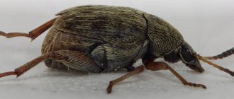
- Sand flea - makes passages, leaving larvae under the skin;
- Tungrate fly - affects open wounds;
- Gadflies.
Myiases are random, facultative and obligate.
Accidental myiasis lesions begin with the entry of fly larvae into healthy organs, usually parasitizing in rotten tissues. This happens when using food and laundry contaminated with insects.
Facultative - the disease develops during the process of infection by larvae, usually living in rotten substances. There are known cases of eggs being laid on unclosed wounds and ulcerative lesions of the skin.
Obligate myiases begin when infected with the larvae of flies - parasites that infect herbivores. The gastrointestinal tract and genitourinary system most often become hostage to parasites. In rare situations, larvae may enter the brain and heart. The most dangerous is intestinal myiasis, which can lead to the death of the patient.
According to the complexity of the disease, myiasis is divided into:
Malignant myiasis. Arthropods leave many larvae on the surface of the infected animal, which penetrate into the body through the eyes and ears with great speed. The brain and internal organs may also be susceptible to infection. Tissues affected by the larvae begin to deteriorate, and the disease progresses to a more severe form with possible death.
Benign myiasis occurs after infection by a single individual. The course of the disease is mild, often unnoticed, and easier treatment is used, with positive results.
Prevention
To prevent the development of myiasis, the following preventive measures must be observed:
- fight flies that appear at home;
- observe the rules of personal hygiene;
- keep the house clean;
- eat only thoroughly washed fruits and vegetables;
- store food correctly (in places inaccessible to insects);
- apply fly repellent;
- promptly treat skin lesions;
- monitor the health of domestic and farm animals, promptly treat skin wounds, and regularly examine for the detection of myiases.
Those who live outside the city need to regularly mow the grass on their property. It is very important that the street toilet, cesspools, and manure heaps are located as far as possible from the house. In order to eliminate the odors emanating from them, which will attract flies, special preparations are used.
Following these rules will help reduce the risk of fly larvae entering the body to zero.
Symptoms of subcutaneous myiasis
The disease always occurs in an acute form. The soft tissues are eaten away by the larvae (damage to the bones is possible). On the outside, thickening of the skin with redness is noticeable. The disease has symptoms similar to intestinal perforation or organ abscess.
Infection with larvae most often occurs through contact with cattle. A female insect (gadfly) can lay eggs on human hair.
The onset of the disease often occurs without pronounced symptoms, later swelling appears as the larvae progress. Then a fistula is formed, in which the larva remains until it matures, after which it leaves the body through the hole.
The disease is accompanied by a severe allergic reaction and symptoms similar to those of toxic poisoning (nausea, vomiting). Symptoms may appear:
- Heat;
- Short-term muscle pain;
- Pain in the heart area;
- A formation under the skin with a sensation of heat and movement under the skin, changing its location;
- General weakness;
- Allergic manifestations, most often urticaria.
Pain during infection can be severe, sometimes leading to fainting. The period of parasitism can last no more than 6 days, but this time is enough to cause serious tissue damage.
Facultative myiases
This group of diseases is caused by flies that normally breed on animal corpses. They cannot lay eggs in a person in the literal sense of the word. Insects lay eggs either in open wounds or on intact skin, ears or nose.
On a note!
Flies do not have an ovipositor that can pierce the skin. Therefore, all photos of a fly laying larvae under the skin are Photoshopped.
The hatched larva itself penetrates the epidermis and develops there for 2-3 weeks. The fully developed maggots leave the body and fall to the ground. There they pupate and after a while adults emerge from the pupae.
Depending on the place where the clutch was made, the larvae penetrate not only under the skin, but also into the eyes, nasal cavities and skull. In this case, the expression “brain eaten” ceases to be figurative. Maggots that have penetrated the cranial cavity feed on brain tissue.
Facultative myiases
If fly larvae get into the eye, they can only be removed surgically. The easiest variant of ophthalmomyasis is the penetration of maggots into the conjunctiva. If it penetrates the eyeball, the disease can result in loss of the organ of vision.
On a note!
In the United States, there have been cases of gray blowfly maggots penetrating deep into the internal tissues of the body.
Removing fly larvae from under the skin, nose and eyes is only possible through surgery.
Treatment
Skin myiasis in a person begins to be treated after an examination and an accurate diagnosis by a medical specialist. The diagnosis is determined by careful examination of the lesions in an enlarged format, when the movement of the larvae is noticeable. This usually takes more than one day. For cutaneous myiasis, the destruction of larvae with medications is not used. Surgical extraction is the most effective method.
At the beginning of treatment, the causative agent of the infection is identified, its location is located, after which the parasites are mechanically removed by the doctor. The lesions are cleaned of larvae by washing the wound with disinfectants (potassium permanganate, furatsilin, etc.). For easier extraction, 2-3 drops of sterile oil are used, as a result of which the parasite, being without air, should appear on the surface and can be easily removed. Treatment of the cavity with disinfectants and the presence of an aseptic dressing are required. To prevent purulent complications, antimicrobial ointments or antibiotics can be used.
It is necessary to use drugs for wound healing. It is also important to use intestinal lavage products and take anthelmintic drugs.
For additional treatment at home, you can prepare an ointment using tar, sulfur and petroleum jelly. Proportions – 6 g sulfur, 1 liter of petroleum jelly, 0.5 liter. tar. All ingredients are mixed and used as an ointment for the affected areas once a day.
Important : the main danger of myiasis is the short period of time from the moment of infection until the beginning of the growth of the larva, which quickly takes root in the patient’s body. Sometimes the size of the tumor reaches 5 cm. In some cases, the speed of movement under the skin is up to several tens of centimeters per day.
Larval development cycle
A fly is an insect with a complete development cycle (complete transformation - holometamorphosis),
which includes the following stages:
- Egg. It takes from 8 to 50 hours to mature, then the formed worm comes out.
- Larva. In the period from 3 to 20 days, fly larvae feed, grow, and molt. After the last molt they turn into a prepupa - the body becomes short and thick. While standing, they find a cool, dry place (they bury themselves in the ground or sand), where they finally pupate. The skin (cuticle) hardens, forming a protective shell - the puparium.
- Doll. A fly is formed inside the puparia within 3-5 days.
- Imago (adult). After 1.5 days, the female is ready for mating.
Under favorable conditions (23-25 ˚C, sufficient food), the fly can live up to 2 months. Larvae, pupae, and fertilized females hibernate at low temperatures, and awaken when warming reaches 10 °C.
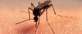
Diseases transmitted by mosquitoes:
• Malaria • Arboviral infections: a. Japanese encephalitis (g.Culex, Aedes) b. Western equine encephalomyelitis (g. Culex, Aedes, Mansonia, Culiseta) c. Eastern equine encephalomyelitis (g. Culiseta, Aedes, Culex, Mansonia) d. Venezuelan equine encephalomyelitis (g. Culex, Aedes, Mansonia, Anopheles) e. St. Louis encephalitis (g. Culex + D.varabilis tick) f. yellow fever (febris fever)(g. Aedes, Haemagogus) g. Dengue fever (dandy fever, date fever, breakbone fever, Aden fever, bouquet fever, Caveat fever, etc.)(g. Aedes) h. Chuqungunia fever (?) (g. Culex, Aedes) i. Tularemia (main - g. Aedes; Anopheles, Culex). • Filariasis: j. Wuchereriasis - helminths Wuchereria bancrofti, W.pacifica - vectors - g.Culex, Aedes, Anopheles k. Brugioses - helminth Brugia malayi - carrier - g.Mansonia, Anopheles
Family Simuliidae.
Black flies - 'midges, midges' - the larvae live only in flowing waters and are demanding of oxygen. Adults are bad fliers. Odagmia ornata Simulium galeratium Simulium argyreatum Schonbaueria pusilla Gnus cholodkovskii Bussodon maculata Simulium morsitans Boopgthora erythrocephala Small blood-sucking insects. Saliva is toxic - causes midge fever - a combination of nausea, headache, fever, swelling of the lymph nodes in sensitive people. In addition, they are carriers and intermediate hosts of a number of human and animal diseases. The main epidemiological significance is oncocerciasis (Onchocerca volvulus). Simuliidae are intermediate hosts in the life cycle of these helminths. Mechanical carriers of tularemia, anthrax, and some hemosporidia.
Family Ceratopogonidae (biting midges)
Biting midges - approximately 5 blood-sucking genera - the main ones are Culicoides, Leptoconops, Forcipomyia, Austroconops. The smallest blood-sucking insects. The larvae live in stagnant waters or damp places - tree hollows, forest litter. Only females feed on blood. They play a significant role in the transmission of microfilariae (in Africa and Latin America - Dipetalonema perstans and D.streptocerca). In southwestern China, Japanese encephalitis virus was isolated from them; in America - Western equine encephalitis virus. They can become infected with tularemia.
Random
The diseases are caused by flies that usually lay eggs in rotting organic matter. A person becomes infected with these larvae accidentally by ingesting them in food or wearing underwear. For the most part, eggs swallowed by a person are dissolved by gastric juice, but sometimes maggots can penetrate the intestines, causing intestinal myiasis. If a fly lays eggs on wet laundry, the larva can penetrate the urethra, causing genitourinary myiasis.
Occasional cavitary myiases are caused by many flies:
- blue meat (Calliphora vicina);
- indoor (Musca domestica);
- green meat (Lucilla sericata);
- brownie (Muscina stabulans);
- small indoor (Fannia canicularis);
- fruit flies (family Drosophilidae);
- cheese (Piophila casei).
These are not the only, but the main types of flies that infect humans.
A person usually causes intestinal myiasis with cheese fly larvae by eating a dubious delicacy: rotten cheese infected with cheese flies.
Parasitology - flies, mosquitoes and other dipterans.
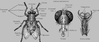
Details Representatives of the class INSECTA (insects) are flies, order DIPTERA (diptera) . There are approximately 120,000 species in the order. The name fly is usually used to refer to insects that have a pair of wings on the mesothorax and a reduced pair of halteres on the metathorax. The halteres are small, club-shaped limbs whose function is a gyroscopic balancing organ. Some parasitic flies have lost their wings for the second time. Flies develop with complete metamorphosis.
Description of the species
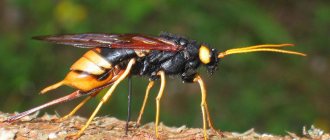
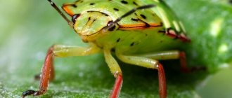
Wohlfahrtiagnifica is a species of gray blowfly. Insects are synanthropic organisms whose lifestyle is closely related to humans. The adult body size is up to 10-14 mm. The main body color is gray. There are three longitudinal black stripes on the mesonotum. There are black spots on the sides and in the upper part of the ovoid abdomen. The body is covered with a light gray coating and stiff hairs. The head is large, large compound eyes of a dark red color. Proboscis of licking type. The antennae are black.
The wings are wide, transparent, well developed. At the base and along the veins there is a smoky color. Jointed black limbs of the running type, covered with bristles. Sexual dimorphism in flies is represented by the location of the eyes. In the photo, the imago of the Wohlfarth fly, judging by the distance between the eyes, is a female; in males it is only 2/3 of the width of the eye.
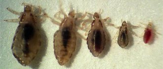
The first instar larvae are small (1.5 mm), worm-shaped, white. After two moults, by the third instar she becomes leaner and larger. The body increases 10 times - up to 15 mm, becomes yellow. There are three oral hooks on the head. The thorax and abdomen segments are covered with brownish spines.
Information. The insect was first described by the German physician Wohlfarth in 1770.
Suborder Nematocera (long-whiskered).
In species of this suborder, the antennae consist of many segments and are thread-like. Initially, they are simple and longer than the head. Wings with a large number of veins, which is a primitive feature. The larvae are active, with a well-developed head capsule; the pupae are often free-swimming. Most larval stages and pupae are aquatic, although some develop in swampy conditions or wet soil.
Family Psychodidae (Phlebotomidae) Subfamily Phlebotominae - sand flies - females feed on plants and blood by thelmophagy; males feed on plants. They fly short distances - up to 1.5 km. Reproduction requires a combination of shade, high humidity, organic waste to feed the larvae, and the presence of dormant hosts. g.Phlebotomus, Sergentomyia, Lutzomyia, Brumtomyia, Warileya Small blood-sucking insects. Saliva is toxic to humans. Specific carriers of papazzi fever (three-day fever), leishmaniasis and bartonellosis (Peruvian fever) (caused by the bacterium Bartonella bacilliformis), and some viral infections.
Family Culicidae Mosquitoes are the most important insect vectors of human diseases and the most common blood-sucking arthropods. Approximately 3,500 species have been described. Mosquitoes (mosquitoes) – complete metamorphosis: egg, larvae, pupa and adult stages. Larvae and pupae can only develop in water. Adult females live about 4-5 months, males - from a week to a month. They fly well - up to 10-30 km.
Subfamily Culicinae
g.Culex – Western equine encephalitis, St. Fox encephalitis viruses. Culex pipiens – house mosquito – vector of Wuchereria bancrofti, Dirofilaria immitis, etc. g.Aedes – yellow fever, tropical fever (epidemic hemorrhagic fever). g.Mansonia, Coquillettidia, Toxorhynchites, Culiseta – filariasis, viruses of western and eastern encephalitis.
Subfamily Anophelinae
g.Bironella, Chagasia – have no medical significance. g.Anopheles - malaria (Plasmodium spp.). Culicidae are specific carriers of 4 species of Plasmodium, three species of filarial nematodes, and many arboviruses (among them the causative agents of yellow fever, Japanese encephalitis, equine encephalomyelitis, and Dengu group viruses). Transmission of these pathogens is possible not only by adult stages, but also transvarially and transphasically.
Why do they appear?
Most often, myiasis occurs in places where people live in unsanitary conditions, where there is no proper body care, there is no normal medical care, and the population neglects hygiene skills.
In most cases, infection occurs due to the negligence and negligence of people, for example, through contact with contaminated objects, feces, soil, as well as through consumption of food or water contaminated by flies.
Human myiases can be caused by various species of dipteran insects, among them:
- Tumbu flies and sand fleas - their females make passages in the thickness of the skin, which become noticeable in the form of painful thickenings. These insects lay eggs, from which larvae then emerge; sometimes their tips peek out and become accessible for removal with tweezers.
- Tungrate flies - love to infect open, weeping wounds or ulcers on the skin, and also cause tissue and cavity forms of the disease (for example, ocular, oral myiasis).
- Gadflies (Russian and sheep) are the cause of subcutaneous, ocular or oral myiasis.
- Sarcophagids (gray blowflies) and cheese flies – can cause disease in the mouth, nose, intestines or skin.
- Calliphorides - they can cause oral myiasis.
- Small housefly - can infect various organs, causing intestinal, genitourinary, ocular or nasal myiasis.
The larvae of these insects have a high degree of vitality, they are not afraid of chemical reagents (acid and alkaline solutions, ethyl alcohol, formaldehyde) and can live for a long time in the intestines and other cavities of the human body, unsuitable for the habitation of adult individuals.