Ear mites in dogs are an external parasite that lives in the outer ear of an animal. If left untreated, infection with ear mites leads to the disease otodectosis with serious consequences. The ectoparasite is widespread in the dog population. The difficulty of dealing with it is that the disease can be transmitted between animals of other species (cats, foxes, ferrets). Dog owners should know what a tick looks like, symptoms and methods of diagnosing otodecosis, control and prevention measures in order to provide timely assistance to their pet. In our material we have collected all the necessary information about this pathology.
What is an ear mite
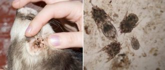
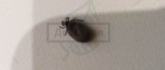
What does a parasite look like? This is a very small insect, not reaching one millimeter, with a greyish-white translucent body, with a gnawing mouthpart. It gnaws through the skin and makes passages under it, laying eggs in them.
Ear mites under a microscope
The wounds become inflamed and fester. The larvae, feeding on the products of suppuration and lymphatic fluid, reach sexual maturity after 3-4 weeks, and then also lay eggs. In a short period of time, an ear mite can damage not only the skin of a dog’s ear, but also the eardrum, and further penetrate into the inner ear and brain.
The disease develops unnoticed - when the animal begins to show signs of the presence of a tick, then, as a rule, a significant part of the ear is affected. In addition, it was noted that the activity of the insect is subject to some cyclicity: a period of activity and bright symptoms is replaced by more or less calm intervals. This may be due to the cyclical development of the parasite. When pathology takes on threatening forms, there are no periods of rest.
Why is it dangerous?
The size of Otodectes cynotis is deceptive. the small parasite leads to death : it gnaws through the eardrum, through which pus enters the inner ear, and from there onto the membranes of the brain. But such a complication is rare and only in emaciated animals that are not treated.
Other possible consequences:
- the addition of a bacterial or fungal infection;
- purulent otitis;
- canal blockage;
- inflammation of the middle or inner ear;
- deafness;
- hematoma - if the pet damages blood vessels when scratching.
How can a dog become infected with ear mites?
Ear mites can be transmitted from one animal to another, regardless of what stage of development it is at. During itching, the dog intensively scratches its ears, contributing to the spread of the parasite throughout the entire surface of the body. That is, the tick and its transitional forms can be located anywhere the pet is.
A healthy animal can “catch” ear mites in the following ways:
- upon contact (even fleeting) with a carrier animal;
- through a grooming item used by an infected dog;
- from a person who had contact with an affected representative;
- through fleas (they can carry tick larvae);
- from the mother (in puppyhood).
Possible complications
In addition to meningitis, the animal may also face other dangerous complications. These include:
- partial or complete deafness;
- dermatitis and allergies;
- purulent otitis;
- weakening of the body's defenses against the background of extensive intoxication;
- secondary infections;
- formation of ear plugs;
- inflammation and rupture of the eardrum.
Remember that puppies and older dogs tolerate the disease much worse than healthy animals. Because of this, they often die from the consequences in a very short time.
Symptoms of ear mites
You can suspect ear mites in a dog if you find a dirty brown mass in the external auditory canal. It is formed from skin scales, particles of the outer integument of shed parasites, and the secretion of the ear glands. All this is mixed with purulent discharge from damaged areas of the epidermis and mite excrement, and leads to severe skin irritation and an inflammatory process.
Other ear mite symptoms:
- hyperemia of the skin of the ear canal;
- severe itching;
- swelling of the ear folds.
The dog is nervous, shakes its head, and often itches. When scratching or shaking the ears, particles of accumulated mass may fly out of the external auditory canal.
Preventive measures
There is no guaranteed way to protect your dog from infection with ear mites. But you can minimize the risk. For this:
- examine the ears once every 2-3 days;
- note the consistency of the plaque;
- observe the behavior of the pet: they are wary when scratching, restlessness, shaking the head;
- Every 4 weeks the dog is treated for blood-sucking parasites, and once every 3 months for helminths;
- do not come into contact with stray dogs and pets whose health they are not sure of;
- Once every 1-2 months, the ears are lubricated with acaricidal preparations for prevention.
A reliable way to protect your dog from otodectosis is to buy an antiparasitic collar with acaricide. It secretes a toxin that is poisonous to ear mites and spreads over the dog’s skin. The accessory is valid for 3-6 months.
The best anti-parasitic collars are considered to be:
- “Preventik” – protects for up to 16 weeks, waterproof, contains fatty acids to improve the condition of the skin and coat, price – 650 rubles;
- "Preventeff" - works for 4 months, does not cause allergies, costs 450 rubles.
Dogs' ears are regularly infested with mites. Any owner will encounter them at least once a year. Therefore, every dog owner is required to know the treatment regimen and the names of acaricidal drugs.
Diagnostics
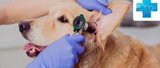
Diagnosing ear mites in dogs is not difficult: during the examination, the veterinarian will take material from the ear and look at it under a microscope. In the chronic form, bacterial seeding of the contents of the ear canal may be required to determine the sensitivity of the insect to drugs and to select the optimal drug. In advanced cases, a specialist may prescribe an X-ray examination or computed tomography to identify the condition of the inner ear and membranes of the brain. Additional diagnostic procedures include: bacterial analysis, scrapings, and allergy tests.
The causative agent of the disease
The causative agent is O. cynotis - a scabies mite-carpet beetle of oval, tortoise-shaped shape, off-white color, with a brown tint in places with stronger chitinization. The head, chest and abdomen are fused into one whole. The proboscis is located on the front of the body. Females are much larger than males. The size of females is 0.32-0.75 mm, males 0.2-0.6 mm. In females, the posterior end is rounded, and in males it is equipped with two abdominal processes with a tuft of setae on each. There are four pairs of legs on the ventral side of the tick. Each leg consists of five segments (coxa, trochanter, femur, tibia, tarsus). At the tops of the tarsi there is a soft membranous pretarsus (sucker or ambulacra). The first three pairs of limbs are well developed, the fourth pair is rudimentary in females. The suckers in females are tulip-shaped, located on short non-segmented rods on the first and second pairs of limbs, in males - on all four. The proboscis is gnawing, horseshoe-shaped. Mites live on the surface of the skin and feed on exfoliated epidermal cells, scales and dry crusts of the skin.
The anal and copulative openings are located at the posterior end of the body. The female lays from several dozen to hundreds of eggs during her life. The eggs are oval in shape, covered with a thin shell, 0.18-0.2 mm long and a maximum width of 0.08-0.09 mm.
Arthropod parasites settle in the hearing organs of a pet, injure delicate skin, and their saliva and feces activate irritation and itching. Insects live only on animals, so people do not become infected.
They prefer to feed on lymph, blood and skin particles.
Is it possible to identify ear mites yourself at home?
There are situations when it is not possible to conduct a microscopic examination of a dog in a clinic. Before treating your pet for otodectosis, you can independently identify the parasite at home. To do this you will need a cotton swab, a piece of dark paper and a magnifying glass. Taking a small amount of plaque from your pet’s external auditory canal with a stick, you need to apply it to paper. If the disease is present, under a magnifying glass you can see moving ticks of a light gray hue.
Important: at the initial stages of pathology development, the population may be small. Therefore, the likelihood that the collected material will contain insects is reduced.
Current tips and tricks
Try on the role of Doctor Aibolit: clean the dog’s ear and examine under a magnifying glass what you manage to pull out of the ear on a piece of black or white paper. A magnifying glass, of course, is not a microscope, but even its capabilities are enough to examine moving microorganisms of light gray color.
If you manage to determine their presence, then you will certainly know that your dog has ticks. If an examination under a magnifying glass does not show anything, this will mean either that the pet is healthy, or that the disease is in the very early stages of enemies - the minimum, and your pet just needs regular prevention of tick infestation - inspection and cleaning of the ears.
Watch your pet carefully. Here are some symptoms (besides furious ear scratching) that should raise your alarm:
- dark brown discharge from the ears,
- skin irritation in the temple area,
- the dog experiences itching in the neck area,
- The dog tilts its head to one side and tries to keep it that way.
If these are your dog’s problems, then you will need the help of a specialist.
Well, you can provide your pet with additional protection - bathe him using insecticidal shampoo. Also, make it a habit to clean your dog’s ears. Purchase a drug from a veterinary pharmacy that softens wax that accumulates in the ear, and use it regularly.
After cleaning your ear, examine the cotton swab: if it is dirty, carry out a similar procedure the next day, and then again and again, until the swab is clean. After this, you can reduce the number of cleanings to one per week.
How to prepare a dog's ear for treatment
Before instilling ear mite drops, you need to clear your pet's ears of any accumulated mass. If the dog resists (not all animals tolerate this procedure stoically, especially if they experience pain), it is better to carry out the procedure together. For small sizes, you can throw a blanket over it or wrap it in a towel. If the pet is large, you should use a muzzle.
During the cleaning process, you must adhere to the following recommendations.
- You need to use sticks, not cotton swabs or disks, as there is a risk of pushing the accumulated mass deeper into the ear canal.
- You should start cleaning from areas located close to the edges of the ear, gradually moving deeper.
- The movements of the wand should be outward.
- If the masses are dry, you can wet the cotton end with peroxide or chlorhexidine. You can't put them in your ear.
- It is advisable to use lotions specifically designed for this purpose to clean your ears.
- If your dog has long hair growing on his ears, it will need to be cut off during treatment.
Hygiene and care - how to rinse your ears before instilling medications
Before instilling medications, the ears must be cleared of accumulated secretions. Dried crusts and scabs are soaked with hydrogen peroxide, furatsilin or herbal infusion (see above). The antiseptic is applied strictly to a cotton swab, since the disc can accidentally push the purulent mass into the sink.
At first, it is better to enlist the help of a friend and put a muzzle on your pet. A small dog can be wrapped in a towel.
While processing, work from the inside out, being careful not to go too deep. Wash your hands thoroughly after each procedure and purchase an Elizabethan collar that prevents scratching of damaged areas.
Prevention
It is impossible to completely prevent your pet from becoming infected with ear mites. However, with the help of preventive measures, you can reduce the likelihood of developing the disease. To do this you need:
- do not allow the dog to come into contact with unfamiliar relatives;
- periodically carefully examine the animal;
- If you find brown plaque in your four-legged friend’s ears, visit the clinic as soon as possible and undergo an examination;
- periodically carry out preventive cleaning with special preparations, which are selected together with a veterinarian, taking into account contraindications and other nuances.
The disease has a favorable prognosis if detected early and treated correctly. At the very beginning of the development of otodectosis, it happens that the ear mites disappear after one cleansing procedure and use of the drug. In advanced cases, you need to be patient, follow the rules of hygiene for your pet’s ears, adhere to the treatment regimen, and increase the dog’s immunity.
FAQ
Can a dog spread ear mites to other pets?
Yes. At risk are dogs, cats, ferrets, and raccoons. All animals in contact must undergo treatment.
Can a person get ear mites from a dog?
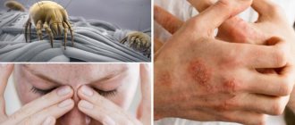
Yes, ticks can move onto the human body, but they only parasitize animals; they quickly die on humans. The risk of infection is minimal; there is no need to be afraid of contact with your pet due to the fact that the tick is transmitted to humans.
However, some people prone to allergic reactions may develop a condition called “false scabies.” The symptoms of this allergic reaction are similar to those of otodectosis. This condition does not require treatment - as soon as the dog is cured, the person will lose all symptoms.
At-risk groups
All dogs, without exception, have a risk of infection. However, there are age groups and certain breeds that are more predisposed to the disease. Most often, puppies and juniors 1.5-6 months old are affected. Long-eared dog breeds have a higher vulnerability. Hunting dogs become infected during hunting from wild animals.
Ear mites are not exclusively a dog disease; they can be transmitted to other animals. If you have other pets, they should be isolated while the sick dog is being treated.


