: 27 Feb 2022, Echo of Memory, volume 85, No. 5/6
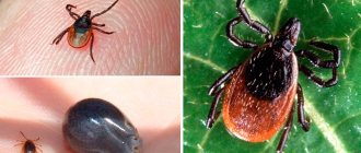
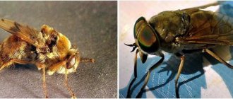
Every year, hundreds of thousands of people visit medical institutions due to a tick bite. For these blood-sucking parasites, humans are only incidental “prey,” but ticks serve as carriers of a whole arsenal of pathogens that cause diseases in humans and animals. Anyone who spends time in nature, including our pets, can face the problem of tick-borne infections; Farm animals also suffer from tick-borne infections. The purpose of this article is to better acquaint readers with the habits and lifestyle of ixodid ticks, primarily common in our country, which can carry about three dozen diseases, including tick-borne encephalitis and borreliosis. In recent years, the problem of tick-borne infections has been aggravated by the fact that the range of these ticks both in Russia and in other countries of the world is rapidly expanding
Ixodid ticks are found in all parts of the world, including islands and coasts of the Arctic and Antarctic, and in various climatic zones - from taiga to desert (Balashov, 1998). Among them there are both fairly “omnivorous” and highly specialized species that feed only on birds, chiropterans or small mammals (Estrada-Pena et al
., 2017).
The main harm from ticks to host animals is associated with the direct parasitism of a large number of ticks on individuals, which can lead to high blood loss or severe intoxication as a result of parasite saliva entering the body. There are known cases of death of wild animals due to massive parasitism of ixodid ticks: wild antelope in the Zimbabwe National Park, young white-tailed deer in the USA, moose in Canada and the northern USA.
The incidence of ticks in farm animals can also be very high in the absence of special anti-tick treatment. Thus, one individual of cattle can simultaneously feed on several dozen, and during the season – several hundred ticks; the total blood loss is calculated in liters! Even cases of death of sheep as a result of several hundred ticks parasitizing them have been described (Balashov, 1998).
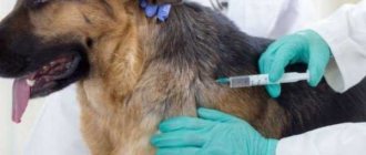
As for humans and their pets, such as dogs, the main danger of ticks for them is the high risk of infection in the event of even a single bite. Ixodid ticks carry pathogens of viral, bacterial and protozoal infections that can infect people. It is impossible to eradicate such diseases, since their foci exist in nature regardless of humans, and the vectors themselves are an integral part of natural ecosystems. And to protect yourself from ticks, you need to know the behavior and ecology of our potential “enemy”.
General description and habitats of ticks
The size of ticks, depending on the variety, can vary from 0.1 to 20 mm. They are distinguished by a solid body, covered on the outside with a cuticle, without pronounced segmentation.
The oral organs of ticks are presented in the form of a cutting, piercing-sucking or gnawing proboscis. Parasites breathe through the entire surface of the body or through the trachea. The digestive system is well developed, but the circulatory system is not sufficiently expressed. One of the characteristic features of ticks is colorless blood. The nervous system is represented by a ganglion and nerve trunks. The excretory system consists of tubes.
When describing ticks, it is worth noting that all species are dioecious. The females of these parasites are mainly oviparous. There are several phases of tick development: egg, larva, which has three pairs of limbs, nymph and adult.
Many ticks (ixodid, argasid, gamasoid) lead an ectoparasitic lifestyle. Moreover, only some belong to endoparasites (pathogens of demodicosis in dogs and cattle).
Some ticks can be stationary parasites for animals (for example, scabies pathogens), while others are only temporary parasites (for example, ixodid ticks).
Ticks most often live in moist, shady places. They can be found not only in forests, parks or gardens, but also in shrubby areas of open spaces, meadows and fields. Ticks are most active in March-May and August-October. In summer, their activity, on the contrary, decreases, and in winter they hibernate. As the ambient temperature rises and air humidity increases, ticks “wake up” in their habitats, and they have an urgent need for food. It is during this period that these parasites actively attack animals and humans.
Duration of parasite activity
Mites of the genus Dermacentor are very cold-resistant. They wake up when the first thawed patches appear. The peak of their activity occurs in April-May: hungry and aggressive adults attack large and medium-sized mammals. By the beginning of summer, the activity of parasites subsides; their summer diapause lasts until August.
In autumn, a second, less strong peak of tick activity is observed. Their life activity ends completely when snow falls.
In autumn, the second active phase of meadow ticks begins, although it is less pronounced than in spring.
The meadow tick is able to survive winter only at the adult stage. Hungry adults go into diapause, and females can be both hungry and well-fed, and males can only be hungry. Nymphs and larvae that do not have time to molt die, regardless of whether they are hungry or full.
Females that have engorged after midsummer enter reproductive diapause. She does not allow them to lay eggs until spring. This process prevents the death of eggs and hatched larvae during the winter cold.
The mechanism of reproductive diapause in female meadow ticks is regulated by the length of daylight hours. This phenomenon is called photoperiodic reaction. The arachnid reacts to the ratio of the length of night and day, and when daylight hours become shorter than a certain period (this value depends on the region), the diapause mechanism is triggered in its body.
Ixodid ticks: external and internal structure, development phases
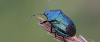
Ixodid ticks are carriers of pathogens that cause piroplasmosis in domestic animals. These ticks can live in forest, steppe, foothill and other zones.
Ixodids are relatively large ticks, the size of which varies from 2 mm to 20 mm, which depends on the condition of the female (hungry females are much smaller than satiated ones). The body appearance of these mites does not have a clear division into the cephalothorax and abdomen. On the outside it is covered with a heterogeneous chitinous layer. On the dorsal and ventral sides of the body there is a large layer of chitin that forms scutes.
The size of the dorsal scute can be used to distinguish a male from a female: in males it occupies the entire upper surface, while in females the scute covers only the anterior part of the body. In the front part of the body are the oral organs, consisting of the proboscis and its base.
The structure of the proboscis of these mites includes the upper and lower jaws, as well as a pair of tentacles. The upper jaws are necessary for cutting the animal's skin before sucking blood. The lower jaw, with teeth pointing backwards, performs the function of fixation. The tentacles are the organ of touch.
As you can see in the photo, depending on the variety, ticks have a long or short proboscis on a quadrangular or hexagonal base:
On the ventral side, at the level of the second pair of legs, there is a genital opening, and behind the fourth pair of limbs there is an anus.
Males have ventral scutes near the anus. On the dorsal side, at the level of the second pair of limbs, some mites have eyes, and at the back of the body there are depressions (festoons). The spiracles (stigmas) are located on the side of the body, behind the fourth pair of legs. There is a plate around them.
One of the structural features of ixodid ticks is the presence in the intestines of a large number of blind outgrowths, which during the period of insect parasitism on animals are filled with blood.
Fertilization of mites occurs on the body of animals. After sucking blood, the females fall away and hide in shelters. After some time, they begin to lay eggs. The total number of eggs laid by one female can vary from 4 to 15 thousand. After this, the females die. Mite eggs are quite large (about 0.5 mm long). They are distinguished by their oval shape and yellow-brown color. The eggs are covered with a hard shell on top.
The internal structure of the ixodid tick is presented in this diagram:
Eggs mature over several weeks (up to a month or more). The appearance of the larva occurs through a crack in the egg shell. The size of the hatched larvae is about 1 mm. She has three pairs of legs. In this case, the larva lacks spiracles, genital opening and peritremes.
To transform into the next stage (nymph), the larva is saturated with the blood of animals. The nymph has four pairs of limbs, but lacks a genital opening.
After being saturated with the blood of animals, the nymph detaches from the host and falls to the ground or turns into an imago directly on the animal’s body. During the complete development of the parasite from the egg to the mature stage, ixodid ticks are saturated with the animal’s blood three times and molt twice. The duration of blood sucking in larvae is on average 5 days, in nymphs it varies from 3 to 10 days, in adults - 10 days. A large number of ticks remain for the winter in the external environment (and at different stages of development).
Depending on the type of development, ixodid ticks can be divided into single-host, two-host and three-host ticks.
All three active stages of development of single-host ticks take place on one animal; only the imago lays eggs in the external environment.
Two-host ticks in the larval and nymphal stages live on one host, and in the adult stage they move to another host.
In the life activity of three-host ticks, a sequential change of three hosts is observed. Moreover, the transition of the parasite from one stage to another always occurs in the external environment. It is this type of development that is most characteristic of ixodid ticks.
Larvae and nymphs of two-host and three-host ticks mainly infect mouse-like rodents; adult ticks and larvae of single-host ticks are more often found on domestic animals.
Trailer ixodids are distinguished by a long proboscis with a quadrangular base. They have no eyes. The anal groove is located anteriorly. In males, the entire ventral surface is covered with scutes. The dorsal shield, limbs and proboscis have a characteristic dark brown tint, and the cuticle of females is grayish-yellow.
The dog tick is one of the most common Ixodid ticks. It is mainly found in the northwestern and central regions of the country, and is absent in the steppe and semi-desert zones. Only one generation of this genus of ticks develops during the year.
Parasites can go hungry for more than two years. In the adult stage, they attack animals in spring and autumn. Dog ticks pose a particular danger to humans, as they transmit diseases such as Lyme disease, encephalitis and tularemia.
Look at the photos of ticks of this variety, the description of which is presented above:
Ticks from the genus Hyalomma (glasseye) are the largest (up to 2.5 cm). They are distinguished by a dark body color, a long proboscis with a quadrangular base and clearly visible eyes. The anal groove is located posteriorly. The males of these ticks are easy to distinguish by their external body structure - they have three pairs of central scutes. These parasites are mainly distributed in steppe, semi-desert and desert zones. Most ticks attack animals in the summer. Representatives of this genus are carriers of causative agents of piroplasmosis, cattle anaplasmosis and many other diseases.
Skin mites are distinguished by a characteristic spotted pattern with a silvery tint on the dorsal shield of males and females. They have a short proboscis with a quadrangular base and eyes. The anal groove is located behind the anus.
Leather mites are widespread in different regions of the country. The main feature of these ticks is the development of parasites according to the three-host type: larvae parasitize mainly mouse-like rodents, nymphs live on larger wild animals, as well as birds, and adults are found on domestic and some large wild animals. Mature ticks mainly parasitize animals in spring and autumn.
These photos illustrate the description of skin-cutting mites:
Bloodsucking mites are relatively small mites that have a short proboscis with a quadrangular base. The anal groove is located behind the anus. Bloodsucking mites lack eyes and ventral scutes in males. These parasites are more common in the steppe and forest-steppe zones, as well as in the foothills. The development of one generation in phases continues for more than a year. Most often, such ticks are carriers of piroplasmosis.
Fan-headed mites are described as relatively small, distinguished by their red-brown color. These parasites are very thermophilic. They have a short proboscis with a hexagonal base, eyes, two pairs of central scutes in males, and a groove located behind the anus. These ticks are most common in the southern, foothill and steppe regions. They parasitize animals in the spring and summer. The development of one generation of fan-headed mites occurs throughout the year.
Most often, two-host and three-host ticks are found, which are carriers of piroplasmid, anaplasmosis and other diseases.
Here you can see photos of ticks, the types of which are described above:
What argas mites look like: appearance (with photo)
Argasid mites (argasids) are external parasites, as well as carriers of some pathogens of infectious and protozoal diseases. They are most often found in the southern regions. Some species of argasids can live in poultry houses and livestock buildings, where they briefly parasitize animals, feeding on their blood at night.
When describing this variety of ticks, it is worth noting that, unlike ixodids, argasid parasites lack dorsal and ventral scutes. The proboscis of imagoes and nymphs is on the ventral surface, so it is not visible from above. The general characteristics of argasid mites are their particularly large size and the presence of a flattened soft body. The proboscis of these parasites is similar in appearance to the proboscis of ixodid ticks.
Nymphs, like adults, have four pairs of limbs, but they lack a genital opening. The round larva has three pairs of limbs. Her proboscis is located in the front of her body.
The body structure of the Persian tick is similar in appearance to a bug. It has a flat, grayish-yellow, ovoid body with a visible edging around it. The length of the tick is 10 mm and the width is 6 mm. The insect has two pairs of legs pointing forward and two pairs pointing back. Ticks go through all stages of development: eggs, larvae, three nymphs and adults. The female parasite is capable of laying from 30 to 100 eggs, from which larvae appear after 2-3 weeks, parasitizing mainly poultry. Each tick in the nymph stage before molting feeds once on the blood of an animal for 1-2 hours, and in the adult stage - several times. It takes 3 to 8 months for the development of one generation of the Persian tick. All stages of the parasite can overwinter. The Persian tick is mainly a carrier of the causative agent of spirochetosis.
The cat tick is distinguished by a thickened body of a grayish color, which is narrowed at the anterior end. Body length can reach 15 mm. The parasite has claws on its feet. The sheep tick parasitizes sheep and other domestic animals.
This parasite goes through all stages of development: eggs, larvae, three nymphs and adults. Female ticks lay eggs in cracks in the walls of barnyards. The entire process can take from 7 to 30 days. During this time, one female is capable of laying from 50 to 500 eggs. After about 30-45 days, larvae emerge from the eggs, which attack animals in the fall. Larvae and nymphs of the cat tick can parasitize animals for up to 45 days. Having had their fill of blood, the nymphs go into hiding, where they turn into adults. Adult ticks repeatedly suck the animal's blood for 10-40 minutes. The main characteristic of cat ticks is winter blood sucking.
In summer, females lay eggs in crevices and cracks in rooms where animals are kept.
Infection with the sheep tick can lead to paralysis (sometimes death) of sheep. In addition, these ticks can be carriers of some sheep hemosporidia. Ticks are carriers and carriers of the pathogens of equine infectious encephalomyelitis, brucellosis and tularemia.
Look what argas mites look like in these photos:
Life cycle
All types of ixodid ticks have a specific life cycle. At each stage, insects pose varying degrees of danger to pets and humans.
Arthropods go through three main stages of development in their lives:
- larvae;
- nymph;
- imago or sexually mature parasite.
The female differs from the male in its smaller size. On the back of the female you can find a shield that covers approximately 2/3 of the entire body. As soon as the female is saturated with blood, her size increases to 11 mm. The individual becomes like a gray pea.
To easily catch prey, the parasite larvae are located on bushes or tall grass with their front legs extended forward. The larvae have the same body shade as mature creatures, but differ in the shade of the cover. A dark, dull color is considered characteristic of them.
The larva can develop into a nymph only if adequate nutrition is available. Once the parasite receives enough blood, it will crawl to the bottom of the forest floor and turn into a nymph, changing its color. Full maturation of the parasite can take from 1 to 1.5 years. Less often, the transformation process drags on for a longer time. The maximum ripening period is limited to 4 years.
Acariform mites
True acariform mites are distinguished by their small body size (from 0.1 to 2 mm). Their mouthparts are of a gnawing or piercing type. There is no circulatory system. These mites go through all stages of development: eggs, larvae, three nymphs and adults. Many acariform mites are the causative agents of scabies in domestic animals. Some of them are involved in the development of ruminant anoplocephalids as intermediate hosts.
Depending on the belonging of scabies pathogens to different genera, it is recommended to establish a diagnosis not for scabies as a whole, but for acarosis, psoroptosis, chorioptosis and notoedrosis.
These photos show what cariform mites look like: